Article
Cryptorchidism in Dogs: Pathophysiology, Diagnosis, and Clinical Relevance
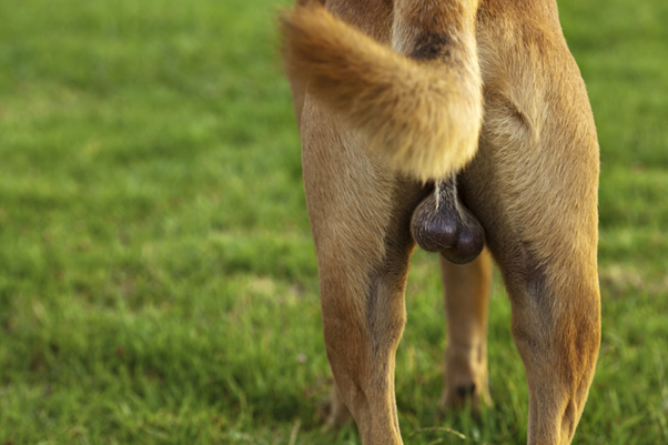
Cryptorchidism, defined as the failure of one or both testes to descend into the scrotum, remains one of the most clinically significant reproductive disorders encountered in small animal practice. Its importance extends beyond a simple developmental anomaly, as it has direct implications for fertility, endocrine function, and oncological risk. Despite its frequency, several aspects of its pathophysiology and long-term consequences continue to challenge veterinary understanding.
Testicular Descent and Developmental Physiology
Testicular descent in the dog is a complex, hormonally regulated process divided into transabdominal, transinguinal, and trans-scrotal phases. Unlike humans, where all phases are completed before birth, dogs complete only the transabdominal phase prenatally. The testes are positioned near the internal inguinal ring at birth, and subsequent descent occurs postnatally, with the transinguinal phase completing shortly after birth and the trans-scrotal phase extending up to 35–140 days postnatally1,2.
This delayed descent has important clinical implications. Dogs are generally considered cryptorchid if the testes are not palpable in the scrotum by 6 months of age, coinciding with closure of the inguinal ring1. The extended window of descent in dogs also introduces variability in diagnosis and highlights the need for careful timing in clinical evaluation.
Hormonal Regulation and Mechanistic Insights
The regulation of testicular descent depends on coordinated hormonal signaling, primarily involving insulin-like factor 3 (INSL3), testosterone, and the hypothalamic–pituitary–gonadal axis. INSL3, produced by fetal Leydig cells, is critical for the transabdominal phase by promoting gubernacular development3. Testosterone and its derivative dihydrotestosterone regulate the transinguinal and trans-scrotal phases through interaction with the genitofemoral nerve, which releases calcitonin gene-related peptide to guide gubernacular migration1.
Disruptions in these hormonal pathways, whether due to genetic mutations or environmental influences, can result in failure of descent. Although anti-Müllerian hormone (AMH) may enhance INSL3 activity, its role remains controversial1,4.
Epidemiology and Clinical Presentation
The reported incidence of cryptorchidism in dogs ranges from 1% to 15%, depending on breed distribution and study design1. In most cases, the retained testis is located within the inguinal canal, although abdominal retention is also observed. Clinically, unilateral cryptorchid dogs may appear normal, whereas bilateral cases are often associated with infertility due to impaired spermatogenesis.
The condition is typically diagnosed through physical examination, but imaging modalities such as ultrasonography are essential in locating abdominal testes. Hormonal assays, particularly AMH, can further assist in confirming the presence of functional testicular tissue.
Pathophysiological Consequences of Cryptorchidism
One of the defining features of cryptorchidism is the exposure of the retained testis to elevated temperatures compared to the scrotal environment. This thermal stress has profound effects on testicular architecture and function. Germ cell depletion is a well-established consequence, primarily mediated by apoptosis induced by heat stress5. Studies in dogs have demonstrated increased lipid peroxidation and upregulation of apoptotic markers such as caspase-3 and caspase-8 in cryptorchid testes1,6.
Interestingly, spermatogonia appear more resistant to thermal injury than spermatocytes and spermatids, leading to selective survival of early germ cells1. This altered cellular environment may contribute not only to infertility but also to neoplastic transformation.
Diagnostic and Clinical Relevance
Diagnosis of cryptorchidism relies on a combination of physical examination, imaging, and hormonal evaluation. However, its clinical significance lies in its association with testicular tumors. Cryptorchid dogs have been reported to have a 9.2 to 13.6 times higher risk of developing testicular neoplasia compared to normal dogs1.
Moreover, tumors in cryptorchid testes tend to develop at an earlier age. This reinforces the importance of early identification and intervention. Orchiectomy remains the recommended treatment, not only to prevent genetic transmission but also to mitigate cancer risk.
Conclusion
Cryptorchidism in dogs represents a multifactorial condition involving developmental, hormonal, and environmental components. Its impact extends beyond reproductive failure, significantly increasing the risk of testicular neoplasia. For practicing veterinarians, early diagnosis and appropriate surgical management are critical in preventing long-term complications and improving patient outcomes.
Reference
- Soto-Heras S, Reinacher L, Wang B, Oh JE, Bunnell M, Park CJ, Hess RA, Ko CJ. Cryptorchidism and testicular cancer in the dog: unresolved questions and challenges in translating insights from human studies. Biology of reproduction. 2024 Aug;111(2):269-91. https://academic.oup.com/biolreprod/article-pdf/111/2/269/58824383/ioae075.pdf
- Gradil C, McCarthy R. Cryptorchidism. In: Monnet E (ed.), Small Animal Soft Tissue Surgery, Second ed. New Jersey: John Wiley & Sons, Inc; 2023: 720–725. https://academic.oup.com/biolreprod/article-pdf/111/2/269/58824383/ioae075.pdf
- Rodprasert W, Virtanen HE, Toppari J. Cryptorchidism and puberty. Front Endocrinol (Lausanne) 2024; 15:1347435. https://www.frontiersin.org/journals/endocrinology/articles/10.3389/fendo.2024.1347435/pdf
- Rey RA. Cryptorchidism is frequently associated with testicular dysfunction. J Endocr Soc 2024; 8:bvae025. https://academic.oup.com/jes/article-pdf/8/4/bvae025/56827948/bvae025.pdf
- Robinson BR, Netherton JK, Ogle RA, Baker MA. Testicular heat stress, a historical perspective and two postulates for why male germ cells are heat sensitive. Biol Rev Camb Philos Soc 2023; 98: 603–622. https://onlinelibrary.wiley.com/doi/pdf/10.1111/brv.12921
- Hernandez-Jardon N, Rojas-Castaneda JC, Landero-Huerta D, Reyes-Cruz E, Reynoso-Robles R, Juarez-Mosqueda MDL, Medrano A, Reyes-Delgado F, Vigueras-Villasenor RM. Cryptorchidism: the dog as a study model. Front Vet Sci 2022; 9:935307. https://www.frontiersin.org/journals/veterinary-science/articles/10.3389/fvets.2022.935307/full
Related Contents

Upcoming Event
CRIs in Anaesthesia Made Easy
Constant Rate Infusions (CRIs) are widely used in veterinary anaesthesia to provide consistent analg...

Upcoming Event
Homeopathy in Pet Animal Practice
Homeopathy continues to be used by some veterinarians and pet owners as a complementary approach in...
.jpg)
Upcoming Event
Advanced Veterinary Transfusion Medicine
Transfusion medicine has become an essential component of modern veterinary critical care and intern...

Article
PRP, IRAP or Stem Cells? Choosing the Right Biologic for Equine Osteoarthritis
Biologics are everywhere—but which one to choose? Regenerative...

Article
Beyond Wear and Tear: Understanding How Osteoarthritis Develops in Performance Horses
For equine athletes, peak performance and joint health exist in a delicate balance. Whether it is a...

Article
Diagnosing Equine Osteoarthritis Earlier: What Every Equine Vet Should Be Using Today
By the time obvious radiographic changes appear, osteoarthritis (OA) has often been active within th...

Article
Beyond NSAIDs: Practical Treatment Strategies for Equine Osteoarthritis
Managing osteoarthritis (OA) in horses is rarely about finding a single "best" treatment....

Article
The Navicular Case That Won't Improve: What Might Be Getting Missed?
For decades, management of podotrochlear syndrome has focused largely on control...