Article
Managing MUO: From First Dose to Long-Term Success
What do you do when the MRI looks clean, but the seizures don’t stop?
That’s the daily puzzle meningoencephalitis of unknown origin (MUO) poses to clinicians.
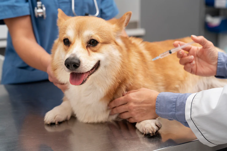
A small-breed dog arrives disoriented, circling, seizuring — you stabilize the patient, but the real question remains: what’s driving the inflammation?
Let’s walk through MUO as it unfolds in real practice from the first presentation to long-term management.
When the Patient Walks in
MUO often strikes without warning — seizures, head tilt, ataxia, or behavior change. The presentation can mimic infection, neoplasia, or intoxication.
Step 1: Stabilize – Control seizures, reduce intracranial pressure, and maintain airway and perfusion.
Step 2: Localize – Conduct a focused neurological exam to determine lesion localization.
Step 3: Prioritize – Exclude hypoglycemia, electrolyte imbalance, or toxins before assuming immune-mediated disease.
If infectious signs are absent, consider early corticosteroid therapy — ideally within five days, as timely immunosuppression has been associated with better outcomes1.
Clinical insight: Act with intent, not haste. Rule out infection, but don’t delay immunotherapy if the clinical pattern fits. Once the patient is stable, the next task is decoding what’s happening within the inflamed CNS.
Connecting the Dots: Diagnostic Reasoning
MUO isn’t a single entity — it encompasses granulomatous (GME), necrotizing (NME), and necrotizing leukoencephalitis (NLE) variants. While each presents subtle differences in imaging and CSF findings, the diagnostic principle remains consistent: rule out infection, confirm inflammation.
Step 4: Image – MRI often reveals multifocal T2/FLAIR hyperintensities.
Step 5: Sample – CSF analysis usually shows mononuclear or lymphocytic pleocytosis.
Step 6: Exclude infection – Perform PCR and serology to rule out infectious etiologies before labeling a case as MUO1.
Common pitfall: Relying solely on MRI.
A patient may have MUO despite a normal MRI if CSF findings indicate inflammation. Nessler & Tipold (2024) highlight the importance of a polythetic approach — interpreting MRI, CSF, and clinical evolution collectively2.
Clinic pearl: Don’t treat the image — treat the inflammation. Once an immune-mediated origin is likely, the focus shifts to achieving durable control.
From Clarity to Control: Treatment in Action
Corticosteroids remain the foundation of MUO therapy, typically prednisolone at 2–4 mg/kg/day, followed by a gradual, deliberate taper over several months.
However, monotherapy may not maintain long-term remission. Adjunct immunosuppressants such as cytarabine, leflunomide, mycophenolate, or cyclosporine can improve disease control and relapse-free survival1.
Step 7: Initiate immunotherapy – Begin corticosteroids once infection has been excluded.
Step 8: Add an adjunct – Introduce one immunosuppressant at a time, allowing for monitoring of efficacy and tolerance.
Step 9: Taper wisely – Reduce corticosteroids slowly, guided by sustained clinical improvement and relapse-free intervals.
Cytarabine infusions (CRI protocols) have shown consistent outcomes and rapid neurological stabilization in clinical use.
A common error is tapering corticosteroids too quickly, which can destabilize immune regulation and precipitate relapse.
Managing the Fallout: Side Effects and Survival
All immunosuppressive protocols carry risk — infections, hepatotoxicity, gastrointestinal upset, or bone marrow suppression. Yet, in practice, relapse remains a greater concern than toxicity.
In a 2023 Scandinavian cohort of 63 MUO dogs, relapse — not drug toxicity — was the leading cause of death3. This emphasizes the need for vigilance and consistent monitoring.
Step 10: Monitor – Perform CBC and serum chemistry every 3–4 weeks during induction, then monthly.
Step 11: Protect – Start gastric protection from day one and educate owners on early infection signs.
Step 12: Communicate – Keep regular follow-ups to ensure adherence and identify relapse early.
Premature tapering remains the most frequent cause of relapse. Clear communication and owner education are essential to maintain control.
Avoiding Clinical Traps
MUO can deceive even experienced clinicians. Some over-rely on imaging; others assume breed predispositions. The most underestimated factor in long-term success is owner compliance.
Step 13: Don’t overtreat the image – MRI normalization does not necessarily mean immune quiescence.
Step 14: Track function, not just findings – CSF stability and neurological recovery are more reliable remission markers1,3.
Step 15: Empower owners – Provide written taper schedules, expected adverse effects, and relapse warning signs.
Empowered owners become effective partners in chronic management — a critical component in preventing relapse and sustaining stability.
Looking Ahead: The Future of MUO Management
Once stability is achieved, the goal shifts from reaction to prevention.
Step 16: Review regularly – Conduct monthly rechecks for six months, then quarterly. MRI or CSF reassessment is indicated only if new neurological signs appear.
Step 17: Rethink immunity – Ongoing research into cytokine modulation and microglial-targeted therapy may refine treatment beyond broad immunosuppression2.
Step 18: Refine goals – Focus on long-term neurological stability and quality of life, not mere survival.
Clinical insight: MUO management has evolved from “suppress and hope” to “monitor, adjust, and anticipate.” The most successful clinicians integrate evolving evidence with compassion — for both patient and owner.
MUO Management in Six Phases — Quick Reference Timeline
Phase 1: Presentation — Stabilize, localize, and rule out emergencies before assuming immune etiology.
Phase 2: Diagnostics — Combine MRI, CSF, and infectious testing; avoid labeling too early.
Phase 3: Induction Therapy — Begin corticosteroids ± adjuncts; taper gradually after achieving stability.
Phase 4: Monitoring — Routine labs, gastric protection, and owner vigilance.
Phase 5: Relapse Prevention — Avoid rapid taper; monitor function and clinical recovery rather than imaging alone.
Phase 6: Future Focus — Explore cytokine and microglial-modulating therapies aimed at immune recalibration.
References
- Beasley MJ, Shores A. Perspectives on pharmacologic strategies in the management of meningoencephalomyelitis of unknown origin in dogs. Frontiers in Veterinary Science. 2023 May 10;10:1167002..
- Nessler JN, Tipold A. Of potential new treatment targets and polythetic approach in meningoencephalitis of unknown origin: a review. Frontiers in veterinary science. 2024 Oct 15;11:1465689.
- Heidemann PL, Erhald B, Koch BC, Gredal H. Investigation of side effects to treatment and cause of death in 63 Scandinavian dogs suffering from meningoencephalitis of unknown origin: a retrospective study. Acta Veterinaria Scandinavica. 2023 Oct 19;65(1):46.
Related Experts
Dr. Hamid Shah
BVSc, MS, PhD PPAM – Pet Practitioners Association of Mumbai
Veterinary
Related Contents

Upcoming Event
CRIs in Anaesthesia Made Easy
Constant Rate Infusions (CRIs) are widely used in veterinary anaesthesia to provide consistent analg...

Upcoming Event
Homeopathy in Pet Animal Practice
Homeopathy continues to be used by some veterinarians and pet owners as a complementary approach in...
.jpg)
Upcoming Event
Advanced Veterinary Transfusion Medicine
Transfusion medicine has become an essential component of modern veterinary critical care and intern...

Article
PRP, IRAP or Stem Cells? Choosing the Right Biologic for Equine Osteoarthritis
Biologics are everywhere—but which one to choose? Regenerative...

Article
Beyond Wear and Tear: Understanding How Osteoarthritis Develops in Performance Horses
For equine athletes, peak performance and joint health exist in a delicate balance. Whether it is a...

Article
Diagnosing Equine Osteoarthritis Earlier: What Every Equine Vet Should Be Using Today
By the time obvious radiographic changes appear, osteoarthritis (OA) has often been active within th...

Article
Beyond NSAIDs: Practical Treatment Strategies for Equine Osteoarthritis
Managing osteoarthritis (OA) in horses is rarely about finding a single "best" treatment....

Article
The Navicular Case That Won't Improve: What Might Be Getting Missed?
For decades, management of podotrochlear syndrome has focused largely on control...