Article
Meniscal Tears in Dogs With Cranial Cruciate Ligament Rupture: Clinical Implications for Practitioners
Introduction
Cranial cruciate ligament (CCL) rupture is a leading cause of stifle instability and hindlimb lameness in dogs. Medial meniscal tears commonly occur alongside complete CCL ruptures, contributing to persistent discomfort, lameness, and joint degeneration. Recognizing the patterns of meniscal injury is essential for veterinarians managing stifle conditions.
Meniscal Function and Injury Mechanism
The stifle menisci provide critical functions, including shock absorption, load distribution, stabilization, and smooth articulation. The medial meniscus, being more firmly attached to the tibial plateau and joint capsule, is more susceptible to injury when the stifle becomes unstable. Following a complete CCL rupture, altered tibiofemoral motion increases stress on the medial meniscus, often leading to tears that require surgical attention1.
Prevalence and Risk Factors
Medial meniscal tears occur frequently in dogs with complete CCL rupture, highlighting the importance of anticipating this complication during evaluation and treatment. Tibial plateau angle (TPA) influences the likelihood of meniscal injury. Dogs with moderate TPA are more prone to meniscal tears, while those with higher TPA (>38°) have a lower risk. The duration of lameness also plays a role: acute lameness is associated with a greater likelihood of medial meniscal injury compared with chronic presentations1.
Clinical Presentation
Dogs with meniscal tears may present with persistent lameness, stifle pain, joint effusion, and difficulty bearing weight. Some dogs exhibit intermittent lameness that worsens with activity. Careful physical examination and attention to subtle signs of instability or pain can help identify affected dogs before surgery1,2.
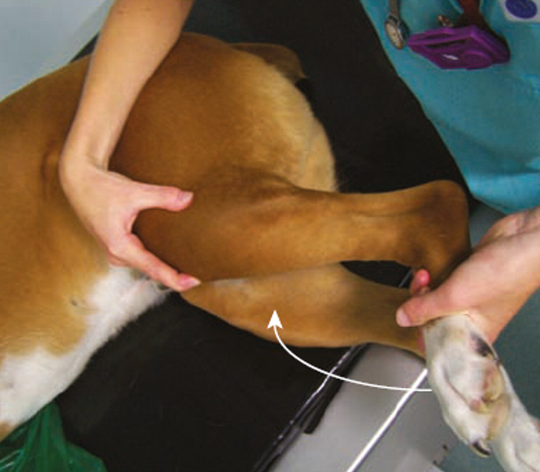
Diagnosis and Assessment
Direct assessment of the meniscus during stifle stabilization is critical. Intra-operative evaluation, including careful probing or arthroscopy, allows identification of meniscal tears and immediate management through partial meniscectomy or other appropriate interventions. Advanced imaging, when available, can support preoperative assessment but does not replace direct visualization1,2.
Management Considerations
Addressing meniscal tears at the time of CCL stabilization is essential to reduce persistent pain and prevent accelerated joint degeneration. Partial meniscectomy is commonly performed for irreparably damaged tissue, while meniscal repair may be considered in cases with sufficient tissue integrity. Postoperative care should include controlled exercise, hydrotherapy, and progressive strengthening to restore limb function and minimize compensatory gait patterns. Weight management is also critical to reduce mechanical stress on the stifle1,2.
Practical Implications
Awareness of meniscal injury patterns, including the influence of TPA and acute lameness, allows veterinarians to plan interventions more effectively. Dogs presenting acutely with complete CCL rupture may require more thorough intra-articular assessment to address medial meniscal tears. Understanding these factors also supports client counseling regarding prognosis and the importance of early intervention1.
Conclusion
Medial meniscal tears are a common and clinically significant complication in dogs with complete CCL rupture. Recognizing risk factors such as tibial plateau angle and acute onset of lameness, combined with careful intra-operative assessment and prompt management, supports improved surgical outcomes, reduces persistent lameness, and helps preserve long-term joint function [1,2].
Reference List
- Bertorelli J, Arnold G, Mertens D. Correlation between the tibial plateau angle and occurrence of medial meniscal tears in dogs with complete cranial cruciate ligament rupture. BMC Vet Res. 2025;21:209.
- Rafla M, Yang P, Mostafa A. Canine Cranial Cruciate Ligament Disease (CCLD): A Concise Review of the Recent Literature. Animals. 2025;15(7):1030.
Related Contents

Upcoming Event
CRIs in Anaesthesia Made Easy
Constant Rate Infusions (CRIs) are widely used in veterinary anaesthesia to provide consistent analg...

Upcoming Event
Homeopathy in Pet Animal Practice
Homeopathy continues to be used by some veterinarians and pet owners as a complementary approach in...
.jpg)
Upcoming Event
Advanced Veterinary Transfusion Medicine
Transfusion medicine has become an essential component of modern veterinary critical care and intern...

Article
PRP, IRAP or Stem Cells? Choosing the Right Biologic for Equine Osteoarthritis
Biologics are everywhere—but which one to choose? Regenerative...

Article
Beyond Wear and Tear: Understanding How Osteoarthritis Develops in Performance Horses
For equine athletes, peak performance and joint health exist in a delicate balance. Whether it is a...

Article
Diagnosing Equine Osteoarthritis Earlier: What Every Equine Vet Should Be Using Today
By the time obvious radiographic changes appear, osteoarthritis (OA) has often been active within th...

Article
Beyond NSAIDs: Practical Treatment Strategies for Equine Osteoarthritis
Managing osteoarthritis (OA) in horses is rarely about finding a single "best" treatment....

Article
The Navicular Case That Won't Improve: What Might Be Getting Missed?
For decades, management of podotrochlear syndrome has focused largely on control...