Article
Diagnostic Precision and Surgical Management of Canine Cryptorchidism: A Clinical Case Approach
Cryptorchidism remains one of the most clinically significant reproductive disorders encountered in small animal practice. While often asymptomatic, its implications, ranging from infertility to neoplasia, demand timely diagnosis and intervention. This case-driven discussion highlights the importance of structured clinical evaluation, imaging, and surgical decision-making in managing abdominal cryptorchidism.
Understanding the Clinical Problem
Cryptorchidism is defined as the failure of one or both testes to descend into the scrotum within the expected developmental window, typically by six months of age1,2. Instead, the testes may remain in the abdominal cavity, inguinal canal, or ectopic locations. The condition is associated with developmental anomalies involving the gubernaculum and suspensory ligaments1.
Clinically, retained testes are often smaller and asymptomatic unless complicated by torsion, inflammation, or neoplasia. Despite the absence of overt pain, hormonal activity persists, contributing to behavioral abnormalities such as hypersexuality and aggression1.
Case Presentation: A Surgical Perspective
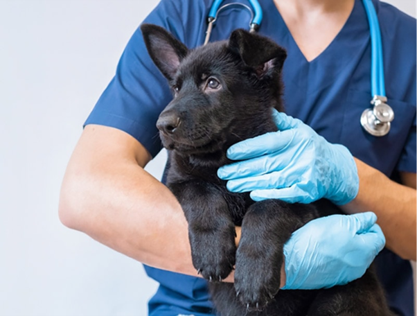
A one-and-a-half-year-old male Golden Retriever weighing 26 kg presented with excessive sexual behavior and abnormal penile protrusion. Physical examination revealed a rudimentary scrotum with the absence of palpable testes. Vital parameters were within normal limits, indicating systemic stability for further diagnostics and intervention.
Palpation, although routinely performed, failed to locate the testes, a limitation observed in nearly 52% of cases1. This reinforces the need for adjunct diagnostic tools in cryptorchid cases.
Role of Imaging in Diagnosis
Radiographic imaging of the abdomen and pelvis did not reveal the retained testes, consistent with known limitations of X-rays in identifying soft tissue structures like undescended testes1.
Ultrasonography, however, proved decisive. Using a trans-abdominal probe (5–10 MHz), both testes were identified based on their echogenicity and structural characteristics. Ultrasound has been shown to have high sensitivity (96.6% for abdominal testes) and is considered superior to exploratory surgery for localization1.
Key ultrasonographic features included:
- Hypoechoic to moderately echogenic oval structures
- Distinct texture compared to surrounding organs
- Absence of normal mediastinum in some cases
This case reinforces ultrasound as a first-line diagnostic modality for non-palpable testes.
Pre-Surgical Evaluation and Planning1
A complete blood profile, including CBC, liver and renal function tests, and glucose levels, revealed no abnormalities. This step is crucial to rule out systemic contraindications before anesthesia and surgery.
The dog was fasted for 12 hours prior to surgery. Premedication included:
- Xylazine (1 mg/kg IM)
- Atropine sulfate (0.02 mg/kg SC)
Prophylactic medications included ceftriaxone (15 mg/kg IV) and meloxicam (0.2 mg/kg). General anesthesia was induced using diazepam and ketamine in a 1:1 ratio.
Surgical Approach and Technique1
A caudal midline laparotomy was performed, extending from the umbilicus to the cranial pubis. This approach provides optimal exposure for abdominal exploration and identification of retained testes.
Both testes were successfully located and excised. The spermatic cord was ligated using absorbable catgut suture material. The abdomen was closed in three standard layers.
Clinical Pearls for Surgeons
- Always explore the caudal abdomen systematically to avoid missing ectopic testes
- Confirm absence of both testes before closure in bilateral cases
- Gentle tissue handling reduces postoperative complications
Postoperative Care
Postoperative management included1:
- Antibiotic therapy for five days
- Daily wound dressing
- Monitoring for complications such as edema, infection, or bleeding
The recovery was uneventful, emphasizing that timely surgical intervention yields excellent outcomes.
Why Early Intervention Matters
Cryptorchid testes carry a significantly higher risk of neoplastic transformation, up to 13.6 times compared to normal testes. Common tumors include seminomas and Sertoli cell tumors, the latter often associated with feminization syndrome1,3.
Additionally, abdominal testes are more prone to torsion due to increased mobility1. Early removal eliminates these risks and prevents long-term endocrine complications.
Take-Home Clinical Insights
- Always suspect cryptorchidism in dogs with absent scrotal testes
- Do not rely solely on palpation, use ultrasonography for confirmation
- Radiography has limited diagnostic value in these cases
- Surgical removal (orchiectomy) remains the gold standard treatment
- Early intervention prevents serious complications like torsion and neoplasia
Conclusion
This case exemplifies the importance of a systematic approach to cryptorchidism—combining clinical suspicion, imaging precision, and surgical expertise. Ultrasonography stands out as a critical diagnostic tool, while timely orchiectomy ensures both therapeutic and preventive benefits. For practicing veterinarians, integrating these steps into routine workflows can significantly improve patient outcomes and reduce long-term complications.
Reference
- Shrestha A, Neupane N, Nepal G, Rimal S. Bilateral Abdominal Cryptorchidism: A Case Study in Dog and Review of Its Relevant Consequences. Mathews Journal of Veterinary Science. 2025 May 30;9(5):1-8. https://www.mathewsopenaccess.com/full-text/bilateral-abdominal-cryptorchidism-a-case-study-in-dog-and-review-of-its-relevant-consequences
- Leslie SW, Sajjad H, Villanueva CA. Cryptorchidism. [Updated 2024 May 5]. In: StatPearls [Internet]. Treasure Island (FL): StatPearls Publishing; 2026 Jan-. Available from: https://www.ncbi.nlm.nih.gov/books/NBK470270/
- Park EJ, Lee SH, Jo YK, Hahn SE, Go DM, Lee SH, Lee BC, Jang G. Coincidence of Persistent Müllerian duct syndrome and testicular tumors in dogs. BMC veterinary research. 2017 Jun 2;13(1):156. https://link.springer.com/content/pdf/10.1186/s12917-017-1068-6.pdf
Related Contents

Upcoming Event
CRIs in Anaesthesia Made Easy
Constant Rate Infusions (CRIs) are widely used in veterinary anaesthesia to provide consistent analg...

Upcoming Event
Homeopathy in Pet Animal Practice
Homeopathy continues to be used by some veterinarians and pet owners as a complementary approach in...
.jpg)
Upcoming Event
Advanced Veterinary Transfusion Medicine
Transfusion medicine has become an essential component of modern veterinary critical care and intern...

Article
PRP, IRAP or Stem Cells? Choosing the Right Biologic for Equine Osteoarthritis
Biologics are everywhere—but which one to choose? Regenerative...

Article
Beyond Wear and Tear: Understanding How Osteoarthritis Develops in Performance Horses
For equine athletes, peak performance and joint health exist in a delicate balance. Whether it is a...

Article
Diagnosing Equine Osteoarthritis Earlier: What Every Equine Vet Should Be Using Today
By the time obvious radiographic changes appear, osteoarthritis (OA) has often been active within th...

Article
Beyond NSAIDs: Practical Treatment Strategies for Equine Osteoarthritis
Managing osteoarthritis (OA) in horses is rarely about finding a single "best" treatment....

Article
The Navicular Case That Won't Improve: What Might Be Getting Missed?
For decades, management of podotrochlear syndrome has focused largely on control...