Article
Clinical Presentation of Demodicosis in Dogs and Cats
Demodicosis presents with a wide spectrum of clinical manifestations in dogs and cats, ranging from mild, localized lesions to severe, generalized disease. Early recognition is essential for appropriate management.
Forms of Demodicosis
Localized Demodicosis
Characterized by:
- One to a few areas of alopecia
- Mild erythema and scaling
- Common involvement of the face and forelimbs
This form is more common in juvenile dogs and may resolve spontaneously in some cases1.
Generalized Demodicosis
Defined by:
- Multiple lesions or widespread involvement
- Pododemodicosis
- Frequent secondary infections
This form typically requires active treatment1.
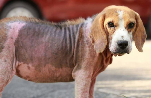
Clinical Signs in Dogs2
Common findings include:
- Patchy or diffuse alopecia
- Erythema
- Scaling and crusting
- Hyperpigmentation in chronic disease
Pruritus is variable and often linked to secondary bacterial infection rather than mite burden1.
Secondary Complications1
Secondary bacterial pyoderma is frequently observed and may present with:
- Papules and pustules
- Crusting
- Draining tracts in severe cases
These complications may increase disease severity and influence treatment decisions.
Clinical Presentation in Cats
Presentation varies by species:
- Demodex cati: Often associated with systemic illness; presents with alopecia and scaling
- Demodex gatoi: Frequently pruritic and may be contagious between cats1
Distribution of Lesions
Common sites include1,2:
- Face and periocular region
- Limbs and paws
- Trunk
Pododemodicosis may be more resistant to treatment and require prolonged therapy.
Clinical Relevance
Recognition of clinical patterns assists in:
- Selecting appropriate diagnostic tests
- Assessing severity
- Guiding treatment decisions
Early identification of generalized disease and complications may improve prognosis.
References
- Mueller RS, Rosenkrantz W, Bensignor E, Karaś‐Tęcza J, Paterson T, Shipstone MA. Diagnosis and treatment of demodicosis in dogs and cats: Clinical consensus guidelines of the World Association for Veterinary Dermatology. Veterinary dermatology. 2020 Feb;31(1):4-e2. https://ebvminpractice.org/wp-content/uploads/2025/09/veterinary-dermatology-2020-mueller-diagnosis-and-treatment-of-demodicosis-in-dogs-and-cats.pdf
- Salem NY, Abdel-Saeed H, Farag HS, Ghandour RA. Canine demodicosis: Hematological and biochemical alterations. Veterinary world. 2020 Jan 10;13(1):68. https://pmc.ncbi.nlm.nih.gov/articles/PMC7020110/pdf/Vetworld-13-68.pdf
Related Contents

Upcoming Event
CRIs in Anaesthesia Made Easy
Constant Rate Infusions (CRIs) are widely used in veterinary anaesthesia to provide consistent analg...

Upcoming Event
Homeopathy in Pet Animal Practice
Homeopathy continues to be used by some veterinarians and pet owners as a complementary approach in...
.jpg)
Upcoming Event
Advanced Veterinary Transfusion Medicine
Transfusion medicine has become an essential component of modern veterinary critical care and intern...

Article
PRP, IRAP or Stem Cells? Choosing the Right Biologic for Equine Osteoarthritis
Biologics are everywhere—but which one to choose? Regenerative...

Article
Beyond Wear and Tear: Understanding How Osteoarthritis Develops in Performance Horses
For equine athletes, peak performance and joint health exist in a delicate balance. Whether it is a...

Article
Diagnosing Equine Osteoarthritis Earlier: What Every Equine Vet Should Be Using Today
By the time obvious radiographic changes appear, osteoarthritis (OA) has often been active within th...

Article
Beyond NSAIDs: Practical Treatment Strategies for Equine Osteoarthritis
Managing osteoarthritis (OA) in horses is rarely about finding a single "best" treatment....

Article
The Navicular Case That Won't Improve: What Might Be Getting Missed?
For decades, management of podotrochlear syndrome has focused largely on control...