Article
Tick-Borne Diseases in Dogs: The Hidden Burden Behind “Healthy” Patients
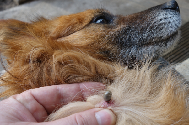
Tick-borne diseases in dogs are not always as obvious as they seem. In many cases, the most challenging patients are the ones that appear completely normal.
In India, canine vector-borne diseases (CVBDs) are highly prevalent due to climatic conditions that favour tick survival and transmission. Common pathogens include Babesia spp., Ehrlichia canis, Anaplasma platys, Hepatozoon canis, and Trypanosoma evansi1. Despite this, a significant proportion of infections remain undiagnosed; especially, in subclinical cases.
The Subclinical Challenge in Everyday Practice
A key concern in clinical settings is the high prevalence of subclinical infections. Dogs may appear healthy during routine examinations, yet harbour haemoparasites.
Microscopy, though widely used, has limitations. Detection depends on adequate parasitemia and observer expertise, making1,2,3,4.
What to do in practice:
- Screen dogs with tick exposure history, even if asymptomatic
- Pay attention to subtle hematological changes
- Repeat testing or escalate diagnostics if suspicion persists
When to Suspect a Tick-Borne Disease
Clinical signs may be mild or non-specific1:
- Intermittent fever
- Lethargy
- Weight loss
- Pale mucous membranes
- Bleeding tendencies
Thrombocytopenia is often a key early finding and may be the only abnormality in some cases5,6.
Clinical tip:
Fever along with thrombocytopenia and tick exposure should always prompt evaluation for tick-borne diseases.
Babesia: A Persistent and Underestimated Threat
Babesia gibsoni is particularly significant in India due to its chronic, relapsing nature and widespread prevalence1. Unlike B. vogeli, it can persist in the host for years, often leading to intermittent clinical signs.
Complete elimination of infection is difficult, and relapses are common despite treatment 1,4.
Clinical tips:
- Monitor PCV and platelet counts during follow-up
- Be prepared for long-term management
- Educate owners about recurrence risk
Co-Infections: The Clinical Complicator
Dogs in endemic areas are frequently exposed to multiple pathogens. Co-infections involving Babesia, Ehrlichia, and Anaplasma are well documented and can significantly alter disease presentation1.
These cases often:
- Present atypically
- Show partial or delayed treatment response
- Have poorer prognosis
Practical approach:
- Suspect co-infection in non-responding cases
- Consider broad diagnostic panels
- Adjust treatment protocols accordingly
Diagnostics: Moving Beyond the Blood Smear
While blood smears remain useful, they are insufficient alone.
Recommended approach:
- CBC and smear for initial screening
- Look for thrombocytopenia, anemia, lymphocytosis
- Use PCR for confirmation and species identification
PCR enables accurate detection and differentiation of species, which is crucial since pathogenicity and treatment response vary1,2,3.
Risk Factors You Should Act On
Several epidemiological factors increase risk1:
- Age: Older dogs show higher infection rates
- Breed: Some breeds may have increased susceptibility
- Environment: Tick-endemic areas with high humidity and temperature
- Stray dog interaction: Community dogs act as reservoirs
In India, the large free-roaming dog population significantly contributes to disease transmission dynamics.
Prevention: A Multi-Layered Strategy
Tick control alone is not enough.
Effective prevention includes1:
- Regular acaricide use (topical/oral/collars)
- Environmental management (kennels, bedding, open areas)
- Routine screening in high-risk dogs
- Owner education on tick checks and early signs
Why This Matters
Subclinical infections are far from harmless. Dogs that appear clinically healthy can act as chronic reservoirs, contributing to ongoing disease transmission. Over time, these infections may lead to long-term complications, particularly affecting the liver and kidneys1,7. Additionally, even asymptomatic dogs can carry high antibody levels and develop chronic inflammatory conditions, reinforcing the need for vigilance in seemingly normal patients1.
The Takeaway for Practice
In clinical practice, it is important not to rely solely on visible symptoms or a single diagnostic test. A negative smear does not always rule out disease, especially in subclinical or chronic cases. Co-infections should always be considered, particularly in endemic regions or in patients with incomplete treatment responses. Proactive screening of high-risk dogs can help in early detection and better disease management. Ultimately, in tick-borne diseases, the absence of symptoms does not equate to the absence of disease.
References
- Vidhya MS, Arunkumar S, Azhahianambi P, KUMAR T, Chandrasekar M. Accurate Diagnosis of Tick-borne Diseases in Working Dogs: The Impact of Unseen Risk Factors. ARCHIVES OF CURRENT RESEARCH INTERNATIONAL Учредители: Sciencedomain International. 2024 Sep 24;24(9):330-40. https://journalacri.com/index.php/ACRI/article/view/897/1898
- Aziz MU, Hussain S, Song B, Ghauri HN, Zeb J, Sparagano OA. Ehrlichiosis in dogs: A comprehensive review about the pathogen and its vectors with emphasis on South and East Asian countries. Veterinary sciences. 2022 Dec 29;10(1):21. https://www.mdpi.com/2306-7381/10/1/21
- Kopparthi J, Chennuru S, Vukka CR, Kumari KN, Prameela DR. Co-infections of major tick-borne pathogens of dogs in Andhra Pradesh, South India. InVeterinary Research Forum 2023 May 15 (Vol. 14, No. 5, p. 295). https://pmc.ncbi.nlm.nih.gov/articles/PMC10278907/pdf/vrf-14-295.pdf
- Karasová M, Tóthová C, Grelová S, Fialkovičová M. The etiology, incidence, pathogenesis, diagnostics, and treatment of canine babesiosis caused by Babesia gibsoni infection. Animals. 2022 Mar 16;12(6):739. https://www.mdpi.com/2076-2615/12/6/739
- Gurjar VS, Singh R, Meena DS, Jeph NK, Choudhary A, Sewag A. Physiological and clinico-hematobiochemical changes in canine babesiosis. Journal of Animal Research. 2023 Apr 1;13(2):303-7. https://www.researchgate.net/profile/Vikram-Gurjar/publication/371401677_Physiological_and_Clinico-Hemato-Biochemical_Changes_in_Canine_Babesiosis/links/65e5ca84c3b52a117015b78b/Physiological-and-Clinico-Hemato-Biochemical-Changes-in-Canine-Babesiosis.pdf
- Miranda EA, Han SW, Rim JM, Cho YK, Yu D, Choi KS, Chae JS. Clinical and subclinical cases of canine babesiosis caused by Babesia gibsoni in the Republic of Korea. Journal of veterinary clinics. 2022;39(5):207-16. https://www.academia.edu/download/99515331/JVC039-05-207.pdf
- Mittal M, Kundu K, Chakravarti S, Mohapatra JK, Nehra K, Sinha VK, Sanjeeth BS, Churamani CP, Kumar A. Canine Monocytic Ehrlichiosis among working dogs of organised kennels in India: A comprehensive analyses of clinico-pathology, serological and molecular epidemiological approach. Preventive veterinary medicine. 2017 Nov 1;147:26-33. https://pmc.ncbi.nlm.nih.gov/articles/PMC7125896/pdf/main.pdf
Related Contents

Upcoming Event
CRIs in Anaesthesia Made Easy
Constant Rate Infusions (CRIs) are widely used in veterinary anaesthesia to provide consistent analg...

Upcoming Event
Homeopathy in Pet Animal Practice
Homeopathy continues to be used by some veterinarians and pet owners as a complementary approach in...
.jpg)
Upcoming Event
Advanced Veterinary Transfusion Medicine
Transfusion medicine has become an essential component of modern veterinary critical care and intern...

Article
PRP, IRAP or Stem Cells? Choosing the Right Biologic for Equine Osteoarthritis
Biologics are everywhere—but which one to choose? Regenerative...

Article
Beyond Wear and Tear: Understanding How Osteoarthritis Develops in Performance Horses
For equine athletes, peak performance and joint health exist in a delicate balance. Whether it is a...

Article
Diagnosing Equine Osteoarthritis Earlier: What Every Equine Vet Should Be Using Today
By the time obvious radiographic changes appear, osteoarthritis (OA) has often been active within th...

Article
Beyond NSAIDs: Practical Treatment Strategies for Equine Osteoarthritis
Managing osteoarthritis (OA) in horses is rarely about finding a single "best" treatment....

Article
The Navicular Case That Won't Improve: What Might Be Getting Missed?
For decades, management of podotrochlear syndrome has focused largely on control...