Article
When Blood Pressure Turns Critical: Recognising Hypertensive Emergency in Small Animals
Arterial blood pressure in mammals is tightly regulated through neurohumoral, myogenic, metabolic, and flow-mediated mechanisms. However, systemic hypertension can develop secondary to various diseases in veterinary patients. In dogs, it is linked to conditions such as acute kidney injury (AKI), chronic kidney disease, diabetes mellitus, immune-mediated haemolytic anaemia, and pheochromocytoma. In cats, chronic kidney disease, hyperthyroidism, and primary hyperaldosteronism are the most common causes1.
Among hypertensive disorders, hypertensive emergency (HE) represents the most severe form, defined by marked elevation in blood pressure with concurrent target organ damage (TOD). Although veterinary thresholds are not standardised, the consequences, damage to the brain, eyes, kidneys, and heart, are well recognised 2.
Acute Clinical Presentation
Hypertensive emergency is typically acute in onset, with clinical signs often developing within a day. This rapid progression makes early recognition essential. In the study, seizures were the most common presenting complaint, emphasising the need to consider systemic hypertension in patients with acute neurological signs1.
Neurological abnormalities frequently included absent menace response, obtundation, anisocoria, and circling, consistent with hypertensive encephalopathy. Ocular findings were also prominent. Fundoscopic examination commonly revealed retinal detachment, followed by retinal haemorrhage. In patients presenting with acute blindness, retinal pathology was often confirmed, although distinguishing between ophthalmic and neurological causes remains important1.
The coexistence of neurological and ocular abnormalities should strongly raise suspicion for hypertensive emergency, particularly in patients with underlying systemic disease.
Target Organ Damage and Pathophysiology
Target organ damage is the defining feature of a hypertensive emergency. In veterinary patients, the most commonly affected organs include the brain, eyes, kidneys, and heart. Among these, neurological and ocular signs are the most clinically apparent2.
Hypertensive encephalopathy results from failure of cerebral autoregulation. The Baylis effect describes vasoconstriction in response to increased pressure, leading to reduced cerebral blood flow and ischaemic injury. In contrast, the “breakthrough theory” suggests that excessive pressure overwhelms autoregulation, resulting in vasodilation, vascular injury, and disruption of the blood–brain barrier. Both mechanisms ultimately lead to vasogenic oedema, a consistent finding in affected patients3.
Blood Pressure Measurement
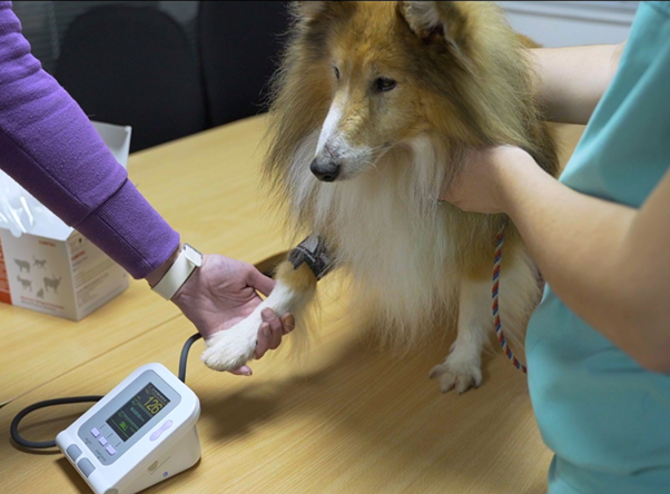
Accurate blood pressure measurement is critical for diagnosis. In this study, all cases were assessed using non-invasive Doppler techniques, following ACVIM guidelines. However, these methods have limitations, and results should be interpreted cautiously as no device fully meets validation standards1,2.
Although invasive arterial blood pressure (IBP) monitoring is the gold standard, it was rarely used, likely due to technical challenges and perceived risks such as infection or haemorrhage. Evidence suggests these risks are generally low1, and IBP should be considered more frequently in critical patients.
The degree of hypertension observed was severe, with dogs showing a median admission SBP of 220 mmHg and cats 230 mmHg, with peaks reaching 300 mmHg.
Diagnostic Workup and Underlying Causes
Identifying the underlying cause is essential for management. Laboratory findings frequently revealed renal dysfunction, with elevated creatinine levels particularly in non-survivors, highlighting the link between hypertension and kidney injury.
AKI was the most common underlying diagnosis in both species. In cats, causes included acute-on-chronic kidney disease, ureteral obstruction, and renal carcinoma. In dogs, AKI was associated with leptospirosis, suspected intoxication, and immune-mediated glomerulonephritis. Other causes included idiopathic hypertension and neoplasia1.
Clinical Relevance
Hypertensive emergency is a true veterinary emergency requiring rapid recognition and intervention. Blood pressure measurement should be routine in patients presenting with seizures, acute blindness, or altered mentation. Neurological and fundoscopic examinations are essential for identifying target organ damage.
In the absence of clear veterinary guidelines, clinicians must rely on clinical judgement and individual patient response. Early detection and timely intervention remain key to improving outcomes in these critically ill patients.
Reference
- Beeston D, Jepson R, Cortellini S. Evaluation of presentation, treatment and outcome in hypertensive emergency in dogs and cats: 15 cases (2003‐2019). Journal of Small Animal Practice. 2022 Oct;63(10):784-91. https://onlinelibrary.wiley.com/doi/pdfdirect/10.1111/jsap.13530
- Acierno MJ, Brown S, Coleman AE, Jepson RE, Papich M, Stepien RL, Syme HM. ACVIM consensus statement: guidelines for the identification, evaluation, and management of systemic hypertension in dogs and cats. Journal of veterinary internal medicine. 2018 Nov;32(6):1803-22. https://academic.oup.com/jvim/article-pdf/32/6/1803/66838718/jvim15331.pdf
- Church ME, Turek BJ, Durham AC. Neuropathology of spontaneous hypertensive encephalopathy in cats. Veterinary pathology. 2019 Sep;56(5):778-82. https://journals.sagepub.com/doi/pdf/10.1177/0300985819849500
Related Contents

Upcoming Event
CRIs in Anaesthesia Made Easy
Constant Rate Infusions (CRIs) are widely used in veterinary anaesthesia to provide consistent analg...

Upcoming Event
Homeopathy in Pet Animal Practice
Homeopathy continues to be used by some veterinarians and pet owners as a complementary approach in...
.jpg)
Upcoming Event
Advanced Veterinary Transfusion Medicine
Transfusion medicine has become an essential component of modern veterinary critical care and intern...

Article
PRP, IRAP or Stem Cells? Choosing the Right Biologic for Equine Osteoarthritis
Biologics are everywhere—but which one to choose? Regenerative...

Article
Beyond Wear and Tear: Understanding How Osteoarthritis Develops in Performance Horses
For equine athletes, peak performance and joint health exist in a delicate balance. Whether it is a...

Article
Diagnosing Equine Osteoarthritis Earlier: What Every Equine Vet Should Be Using Today
By the time obvious radiographic changes appear, osteoarthritis (OA) has often been active within th...

Article
Beyond NSAIDs: Practical Treatment Strategies for Equine Osteoarthritis
Managing osteoarthritis (OA) in horses is rarely about finding a single "best" treatment....

Article
The Navicular Case That Won't Improve: What Might Be Getting Missed?
For decades, management of podotrochlear syndrome has focused largely on control...