Article
From Scratch to Solution: A Chairside Guide to Managing Canine Atopic Dermatitis
Canine atopic dermatitis (cAD) is a chronic, inflammatory skin condition affecting up to 30% of dogs1. For Indian veterinary practice, understanding immunopathogenesis and evidence-based, locally applicable therapies is essential for effective management.
Recognizing the Clinical Patterns
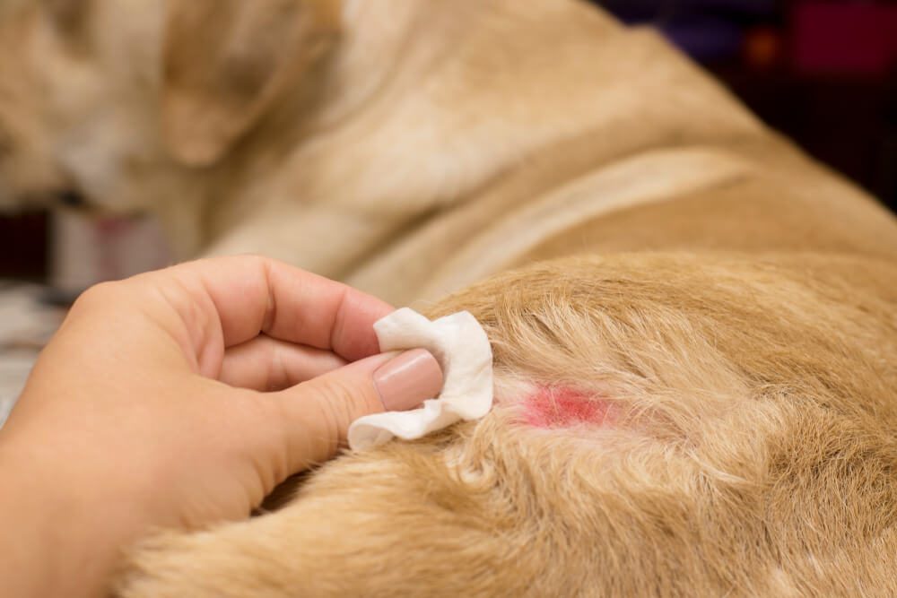
Dogs with cAD present with persistent pruritus, erythema, lichenification, and frequent secondary bacterial or fungal infections2,3. Self-induced alopecia from scratching or licking is common, and lesion severity varies across patients4. The disease arises from genetic predisposition, environmental triggers, and immune dysregulation5,6.
Immunopathogenesis in Practice
Previously considered a Th2-dominated disorder, cAD is now understood as multi-axial. Acute lesions typically show Th2 cytokines (IL-4, IL-13), while chronic lesions involve Th1 (IFN-γ, IL-12) and Th22 (IL-22) responses1,5. Other mediators, including IL-7, IL-34, IL-36, macrophage migration inhibitory factor (MIF), and neurogenic factors (NGF, substance P, periostin), further drive inflammation7. Recognizing these pathways guides targeted therapy.
Targeted and Indirect Anti-Cytokine Therapies Available in India
- JAK inhibitors
- Oclacitinib selectively inhibits JAK1, primarily blocking IL-31, IL-4, and IL-13, reducing pruritus rapidly. Recommended dosing: 0.4–0.6 mg/kg orally twice daily for up to 14 days, then once daily for maintenance. Contraindicated in dogs <12 months or <3 kg, or with immunosuppression or progressive neoplasia1,8. Rapid onset makes it ideal for pruritus-dominant phenotypes.
- Calcineurin inhibitors
- Ciclosporin binds cyclophilin to inhibit calcineurin, suppressing IL-2, IL-4, and IFN-γ. Oral administration at ~5 mg/kg daily effectively reduces pruritus and lesion severity, especially in chronic cAD, with better long-term tolerability than systemic glucocorticoids. Contraindicated in dogs <6 months or <2 kg, or with neoplasia; caution in diabetic patients1,9.
- Tacrolimus inhibits calcineurin locally and is useful for localized lesions (footpads, skin folds, peri-aural regions), particularly when systemic therapy is contraindicated or to minimize systemic immunosuppression1.
- Monoclonal antibodies
- Anti-IL-31 monoclonal antibody targets IL-31 specifically, providing rapid pruritus relief within 8–12 hours after subcutaneous injection. Dosing is every 4–8 weeks, depending on weight. Its highly selective mechanism minimizes systemic immunosuppression, making it safe for dogs with comorbidities or during vaccination schedules10,11,12.
Chairside Strategy for Indian Practice
- Rapid pruritus control: Oclacitinib or anti-IL-31 monoclonal antibody depending on phenotype, owner compliance, and convenience.
- Long-term management: Ciclosporin or anti-IL-31 monoclonal antibody are effective and safe.
- Phenotype-guided therapy:
- Pruritus-dominant: anti-IL-31 mAb or oclacitinib
- Chronic/mixed inflammatory patterns: oclacitinib or ciclosporin
- Localized lesions: tacrolimus ointment
- Monitoring: Use PVAS and CADESI scores to track response and adjust dosing.
Integrating Precision Medicine
Combining symptomatic pharmacotherapy with allergen control, infection management, and epidermal barrier care improves outcomes and reduces relapses. Breed-specific differences, cytokine-phenotype variation, and pharmacokinetics highlight the importance of individualized protocols1.
Looking Ahead
Future cAD management will focus on selective cytokine blockade without disrupting systemic immune homeostasis. Using these therapies appropriately allows veterinarians to achieve rapid relief, reduce flares, and minimize adverse effects—a practical, chairside roadmap from “scratch to solution.”
References
- Wichtowska A, Olejnik M. Anti-Cytokine Drugs in the Treatment of Canine Atopic Dermatitis. International Journal of Molecular Sciences. 2025 Nov 13;26(22):10990. https://doi.org/10.3390/ijms262210990
- Brunner PM, Guttman-Yassky E, Leung DY. The immunology of atopic dermatitis and its reversibility with broad-spectrum and targeted therapies. Journal of Allergy and Clinical Immunology. 2017 Apr 1;139(4):S65-76. https://www.jacionline.org/article/S0091-6749%2817%2930205-1/pdf
- Outerbridge CA, Jordan TJ. Current knowledge on canine atopic dermatitis: Pathogenesis and treatment. Advances in small animal care. 2021 Nov 1;2:101-15. https://pmc.ncbi.nlm.nih.gov/articles/PMC9204668/pdf/nihms-1810001.pdf
- Hensel P, Santoro D, Favrot C, Hill P, Griffin C. Canine atopic dermatitis: detailed guidelines for diagnosis and allergen identification. BMC veterinary research. 2015 Aug 11;11(1):196. https://link.springer.com/content/pdf/10.1186/s12917-015-0515-5.pdf
- Drechsler Y, Dong C, Clark DE, Kaur G. Canine atopic dermatitis: prevalence, impact, and management strategies. Veterinary Medicine: Research and Reports. 2024 Dec 31:15-29. https://www.tandfonline.com/doi/pdf/10.2147/vmrr.s412570
- Hensel P, Saridomichelakis M, Eisenschenk M, Tamamoto‐Mochizuki C, Pucheu‐Haston C, Santoro D, International Committee on Allergic Diseases of Animals (ICADA), Banovic F, Bensignor E, DeBoer D, Griffin C. Update on the role of genetic factors, environmental factors and allergens in canine atopic dermatitis. Veterinary Dermatology. 2024 Feb;35(1):15-24. https://onlinelibrary.wiley.com/doi/pdf/10.1111/vde.13210
- Pucheu‐Haston CM, Bizikova P, Marsella R, Santoro D, Nuttall T, Eisenschenk MN. Lymphocytes, cytokines, chemokines and the T‐helper 1–T‐helper 2 balance in canine atopic dermatitis. Veterinary dermatology. 2015 Apr;26(2):124-e32. https://www.research.ed.ac.uk/files/19114604/ICADA_AD_review_lymphocytes_cytokines_chemokines_and_the_TH1_TH2_balance.pdf
- Cosgrove SB, Cleaver DM, King VL, Gilmer AR, Daniels AE, Wren JA, Stegemann MR. Long‐term compassionate use of oclacitinib in dogs with atopic and allergic skin disease: safety, efficacy and quality of life. Veterinary Dermatology. 2015 Jun;26(3):171-e35. https://onlinelibrary.wiley.com/doi/pdfdirect/10.1111/vde.12194
- Olivry T, DeBoer DJ, Favrot C, Jackson HA, Mueller RS, Nuttall T, Prélaud P, International Committee on Allergic Diseases of Animals. Treatment of canine atopic dermatitis: 2015 updated guidelines from the International Committee on Allergic Diseases of Animals (ICADA). BMC veterinary research. 2015 Aug 16;11(1):210. https://link.springer.com/content/pdf/10.1186/s12917-015-0514-6.pdf
- Moyaert H, Van Brussel L, Borowski S, Escalada M, Mahabir SP, Walters RR, Stegemann MR. A blinded, randomized clinical trial evaluating the efficacy and safety of lokivetmab compared to ciclosporin in client‐owned dogs with atopic dermatitis. Veterinary dermatology. 2017 Dec;28(6):593-e145. https://onlinelibrary.wiley.com/doi/pdfdirect/10.1111/vde.12478
- Marsella R, Ahrens K, Wilkes R, Trujillo A, Dorr M. Comparison of various treatment options for canine atopic dermatitis: a blinded, randomized, controlled study in a colony of research atopic beagle dogs. Veterinary dermatology. 2020 Aug;31(4):284-e69. https://www.academia.edu/download/91542663/vde.1284920220925-1-1lufvkx.pdf
- Cheung BY. In dogs with atopic skin disease, is lokivetmab more effective than oclacitinib in reducing the score of a recognised scoring system?. Veterinary Evidence. 2022 Jun 22;7(2). https://veterinaryevidence.org/index.php/ve/article/download/569/757
Related Contents

Upcoming Event
CRIs in Anaesthesia Made Easy
Constant Rate Infusions (CRIs) are widely used in veterinary anaesthesia to provide consistent analg...

Upcoming Event
Homeopathy in Pet Animal Practice
Homeopathy continues to be used by some veterinarians and pet owners as a complementary approach in...
.jpg)
Upcoming Event
Advanced Veterinary Transfusion Medicine
Transfusion medicine has become an essential component of modern veterinary critical care and intern...

Article
PRP, IRAP or Stem Cells? Choosing the Right Biologic for Equine Osteoarthritis
Biologics are everywhere—but which one to choose? Regenerative...

Article
Beyond Wear and Tear: Understanding How Osteoarthritis Develops in Performance Horses
For equine athletes, peak performance and joint health exist in a delicate balance. Whether it is a...

Article
Diagnosing Equine Osteoarthritis Earlier: What Every Equine Vet Should Be Using Today
By the time obvious radiographic changes appear, osteoarthritis (OA) has often been active within th...

Article
Beyond NSAIDs: Practical Treatment Strategies for Equine Osteoarthritis
Managing osteoarthritis (OA) in horses is rarely about finding a single "best" treatment....

Article
The Navicular Case That Won't Improve: What Might Be Getting Missed?
For decades, management of podotrochlear syndrome has focused largely on control...