Article
Hindlimb Lameness in Dogs - Differential Diagnosis & Field Management
Hindlimb lameness is a frequent presenting complaint in Indian veterinary clinics, especially among large-breed dogs. Accurate diagnosis and effective field management of this issue require a structured approach to differentiating common orthopedic conditions such as cranial cruciate ligament rupture (CCLR), hip dysplasia, patellar luxation, and musculoskeletal trauma.
Cranial cruciate ligament rupture is the leading cause of hindlimb lameness in adult dogs. It is commonly seen in Labradors, German Shepherds, and Indian Indies. Dogs typically present with acute, non-weight-bearing lameness and stifle joint effusion. Key diagnostic tests include the cranial drawer and tibial thrust tests. Radiographs may reveal joint effusion and osteophytes in chronic cases (Duerr et al., 2016).
Hip dysplasia, often bilateral and genetically predisposed, affects the coxofemoral joint and presents as bunny-hopping gait, difficulty rising, and muscle wasting. Diagnosis requires sedation and proper radiographic positioning (extended VD view). The Ortolani sign can be helpful in detecting hip laxity in young dogs.
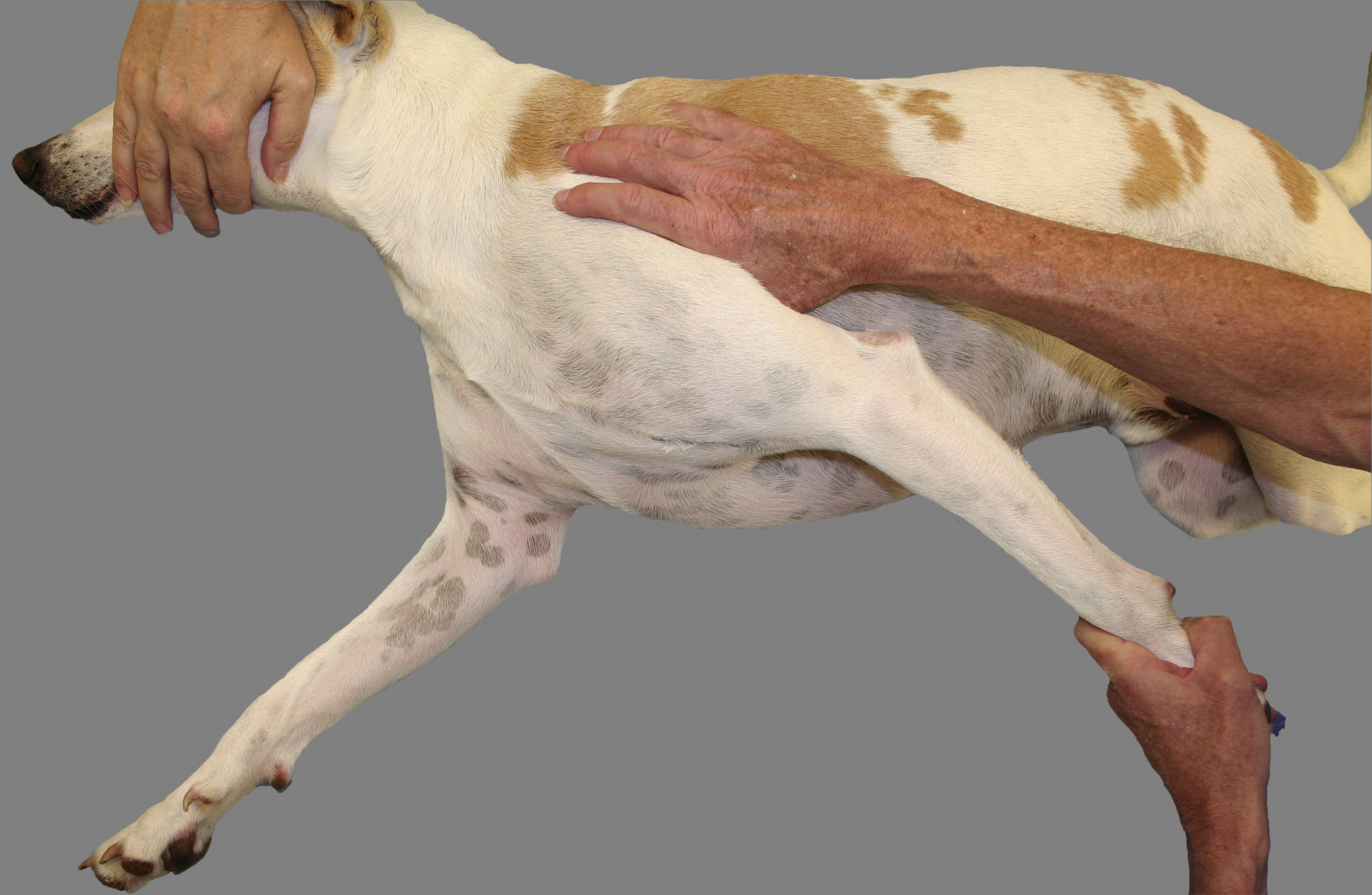
Patellar luxation, particularly medial luxation, is more common in small breeds but can be seen in medium-breed Indian dogs. Clinical signs include intermittent skipping lameness and reluctance to jump. Diagnosis is manual palpation often reveals spontaneous luxation
during flexion (Innes et al., 2000).
In field setups with limited imaging access, a tiered management approach is essential(Di Dona et al., 2018):
- Start with cage rest (5–7 days), cold compress, and NSAIDs (e.g., carprofen 4.4 mg/kg SID).
- Monitor response and re-evaluate lameness at day 7–10.
- Persistent or non-improving cases should be referred for orthopedic consultation and imaging.
Rehabilitation exercises such as assisted walking, weight shifts, and underwater treadmill therapy improve recovery post-diagnosis or surgery. Owner education on exercise restriction, dietary control, and joint supplementation (e.g., omega-3s, glucosamine) is crucial to avoid recurrence.
References:
- Duerr FM et al. (2016). https://pubmed.ncbi.nlm.nih.gov/25328024/
- Innes JF & Bacon D. (2000). https://pubmed.ncbi.nlm.nih.gov/11058021
- Di Dona, F., Della Valle, G., & Fatone, G. (2018). https://doi.org/10.2147/vmrr.s142545
Related Contents

Upcoming Event
CRIs in Anaesthesia Made Easy
Constant Rate Infusions (CRIs) are widely used in veterinary anaesthesia to provide consistent analg...

Upcoming Event
Homeopathy in Pet Animal Practice
Homeopathy continues to be used by some veterinarians and pet owners as a complementary approach in...
.jpg)
Upcoming Event
Advanced Veterinary Transfusion Medicine
Transfusion medicine has become an essential component of modern veterinary critical care and intern...

Article
PRP, IRAP or Stem Cells? Choosing the Right Biologic for Equine Osteoarthritis
Biologics are everywhere—but which one to choose? Regenerative...

Article
Beyond Wear and Tear: Understanding How Osteoarthritis Develops in Performance Horses
For equine athletes, peak performance and joint health exist in a delicate balance. Whether it is a...

Article
Diagnosing Equine Osteoarthritis Earlier: What Every Equine Vet Should Be Using Today
By the time obvious radiographic changes appear, osteoarthritis (OA) has often been active within th...

Article
Beyond NSAIDs: Practical Treatment Strategies for Equine Osteoarthritis
Managing osteoarthritis (OA) in horses is rarely about finding a single "best" treatment....

Article
The Navicular Case That Won't Improve: What Might Be Getting Missed?
For decades, management of podotrochlear syndrome has focused largely on control...