Article
Diagnostic Approach to the Icteric Cat: Avoiding Pitfalls and Making Clinical Decisions
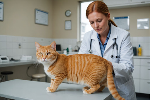
Icterus in feline patients often presents as a visually striking yet diagnostically complex condition. While the yellow discoloration of tissues is unmistakable, identifying the underlying cause requires a systematic and clinically informed approach. Misinterpretation of laboratory values or over-reliance on single parameters can lead to diagnostic errors. Therefore, a structured diagnostic strategy is essential for effective case management.
Initial Clinical Assessment: More Than Just Yellow Discoloration
The evaluation of an icteric cat begins with a thorough clinical examination and history. Clinical signs such as lethargy, anorexia, vomiting, weight loss, and abdominal pain are commonly observed across all types of icterus. However, subtle differences may provide diagnostic clues1.
For example, anemia and dark urine may suggest a hemolytic origin, while pale stools and pruritus are more indicative of obstructive pathology. However, these signs are not definitive and should always be interpreted in conjunction with laboratory findings1.
Approach to Prehepatic Icterus
The first step in any icteric case is to rule out hemolysis. Routine hematological analysis, including packed cell volume (PCV) and complete blood count (CBC), can quickly identify anemia. Blood smear examination remains indispensable for detecting morphological changes such as polychromasia, anisocytosis, and spherocytosis, which indicate regenerative anemia2.
Reticulocyte counts further help assess the regenerative response, although clinicians must remember that regeneration may not be evident in the early stages of disease (first 3–4 days). Additional findings such as hemoglobinemia, hemoglobinuria, and autoagglutination support a diagnosis of hemolytic anemia.
A critical clinical insight is that bilirubin levels in pure hemolytic icterus rarely exceed 3–4 mg/dL. Values beyond this threshold should prompt investigation into hepatic or posthepatic causes1.
Common Diagnostic Pitfalls
One of the most significant pitfalls in diagnosing icterus is the reliance on conjugated versus unconjugated bilirubin levels. While traditionally used to differentiate types of icterus, this distinction is not reliable in clinical practice. In hemolytic conditions, unconjugated bilirubin may predominate initially, but conjugated bilirubin can increase over time, leading to confusion.
Similarly, elevated alanine aminotransferase (ALT) levels should not be immediately attributed to primary liver disease. Severe hemolytic anemia can cause hypoxia-induced hepatocellular injury, resulting in increased ALT levels1. Recognizing these nuances is crucial to avoid misdiagnosis.
Evaluating Hepatic Causes
If hemolysis is ruled out, attention shifts to hepatic causes. Liver enzyme analysis, including ALT, AST, and ALP, provides valuable information regarding hepatocellular injury and cholestasis. However, liver disease does not always produce marked hyperbilirubinemia, and mild to moderate increases are more typical.
A detailed history is particularly important in this context. Exposure to hepatotoxic drugs or toxins, recent infections, and dietary changes can provide critical diagnostic clues. Serological testing for infectious diseases such as FIP and toxoplasmosis may also be warranted1.
Role of Imaging in Posthepatic Icterus
When both hemolytic and primary hepatic causes are excluded, posthepatic icterus must be considered. Ultrasonography is the most valuable diagnostic tool in such cases. In normal cats, intrahepatic bile ducts are not visible, and the common bile duct measures less than 4 mm in diameter.
Dilation of bile ducts or the biliary tree is a key indicator of obstruction. Additionally, ultrasound can identify mass lesions, gallbladder abnormalities, and pancreatic pathology, which are common causes of extrahepatic bile duct obstruction3.
In posthepatic icterus, bilirubin levels can rise dramatically, often exceeding 20 mg/dL, providing another distinguishing feature1.
Integrating Laboratory and Clinical Data
Effective diagnosis of icterus relies on integrating multiple data points rather than focusing on a single parameter. Hematology, biochemistry, imaging, and clinical findings must be interpreted collectively. For instance, a cat with mild anemia, elevated ALT, and significantly high bilirubin may have a combination of hemolytic and hepatic pathology.
Conclusion
Diagnosing icterus in cats is a nuanced process that requires both scientific understanding and clinical judgment. Avoiding common pitfalls, such as over-reliance on bilirubin fractions or misinterpretation of liver enzymes, is essential for accurate diagnosis. By adopting a systematic and integrative approach, veterinarians can effectively identify the underlying cause of icterus and initiate appropriate treatment strategies.
Reference
- ÖZCAN AC, AKTAS MS. Icterus in Cats. Turkish Journal of Veterinary Internal Medicine. 2024;3(2):12-21. https://dergi.veterinerichastaliklari.org/index.php/vihder/article/download/35/32
- Huang L, Qian X, Wu C, Liu P, Zhao Y, Xu Z, Liu P. Diagnosis And Treatment Of A Cat With Immune Mediated Hemolytic Anemia (Imha). https://agrobiologicalrecords.com/articles/2-4-Vol-6-7-12-2021-ABR-21-0367.pdf
- Griffin MA, Culp WT, Giuffrida MA, Selmic LE, Denitz JC, Perry JA, Schoelkopf AC, Milovancev M, Phillips H, Wallace ML, Steffey MA. Choledochal stenting for treatment of extrahepatic biliary obstruction in cats. Journal of veterinary internal medicine. 2021 Nov;35(6):2722-9. https://onlinelibrary.wiley.com/doi/pdf/10.1111/jvim.16176
Related Contents

Upcoming Event
CRIs in Anaesthesia Made Easy
Constant Rate Infusions (CRIs) are widely used in veterinary anaesthesia to provide consistent analg...

Upcoming Event
Homeopathy in Pet Animal Practice
Homeopathy continues to be used by some veterinarians and pet owners as a complementary approach in...
.jpg)
Upcoming Event
Advanced Veterinary Transfusion Medicine
Transfusion medicine has become an essential component of modern veterinary critical care and intern...

Article
PRP, IRAP or Stem Cells? Choosing the Right Biologic for Equine Osteoarthritis
Biologics are everywhere—but which one to choose? Regenerative...

Article
Beyond Wear and Tear: Understanding How Osteoarthritis Develops in Performance Horses
For equine athletes, peak performance and joint health exist in a delicate balance. Whether it is a...

Article
Diagnosing Equine Osteoarthritis Earlier: What Every Equine Vet Should Be Using Today
By the time obvious radiographic changes appear, osteoarthritis (OA) has often been active within th...

Article
Beyond NSAIDs: Practical Treatment Strategies for Equine Osteoarthritis
Managing osteoarthritis (OA) in horses is rarely about finding a single "best" treatment....

Article
The Navicular Case That Won't Improve: What Might Be Getting Missed?
For decades, management of podotrochlear syndrome has focused largely on control...