
Article
Radiography in Canine Osteoarthritis: Current Role, Strengths, and Limitations
Canine osteoarthritis (OA) is one of the most frequently encountered orthopedic conditions in small animal practice and remains a major cause of chronic pain, reduced mobility, and diminished quality of life1,2,3. Although advances in diagnostic imaging have expanded the ability to evaluate joints in greater detail, radiography continues to be the primary imaging modality for diagnosing OA in everyday veterinary practice. Its widespread availability, familiarity, and cost-effectiveness make it an indispensable tool for assessing dogs with suspected degenerative joint disease.
Understanding both the capabilities and the limitations of radiography is essential for interpreting findings appropriately and making informed clinical decisions.
Recognizing Radiographic Features of Osteoarthritis
The integration of radiography into veterinary practice transformed the diagnosis of OA by allowing clinicians to identify structural joint changes in living patients rather than relying solely on clinical examination. The characteristic radiographic features recognized today have remained largely consistent over time and include:
- Periarticular osteophyte formation
- Subchondral sclerosis
- Joint remodeling
- Joint effusion or soft tissue swelling
- Joint space narrowing (JSN), although less consistently evaluated in dogs than in human medicine1,4
These changes reflect structural adaptation of the joint as OA progresses and provide valuable evidence supporting the diagnosis. Radiographic scoring systems have also been developed to document disease severity and monitor progression over time1,4.
Why Radiography Remains the First-Line Imaging Modality
Despite the availability of advanced imaging techniques, radiography remains the standard imaging approach for canine OA because it is inexpensive, widely accessible, well established, and safe1. For most first-opinion practices, it continues to provide the foundation for diagnosing degenerative joint disease and evaluating long-term progression.
Radiographs are particularly useful for identifying bony remodeling associated with established OA. Osteophytes, sclerosis, joint remodeling, and other skeletal changes can often be detected with confidence, allowing clinicians to confirm the presence of chronic joint disease and support treatment planning.
Its accessibility also makes serial imaging practical when monitoring disease progression or evaluating response to management over time.
Understanding the Limitations
While radiography remains invaluable, it is equally important to recognize what it cannot demonstrate.
Radiographic severity does not always reflect the degree of pain or functional impairment experienced by the patient1,5. Dogs with marked radiographic changes may show relatively mild clinical signs, whereas others with limited radiographic abnormalities may experience significant lameness or discomfort. Clinical assessment therefore remains essential when interpreting imaging findings.
Another important limitation is that radiographs primarily depict bone. Because soft tissues, including articular cartilage, synovial fluid, and synovium, have similar radiographic opacity, they cannot be differentiated reliably on standard radiographs1. As a result, many early pathological changes occurring within the joint remain undetected until secondary bony remodeling develops.
Radiography is therefore more effective at identifying established OA than detecting the earliest stages of disease, when structural changes may still be subtle.
Practical Clinical Insights
For practicing veterinarians, radiographic findings should always be interpreted alongside the patient's history, orthopedic examination, gait assessment, and overall clinical presentation.
Keep the following points in mind:
- Use radiographs to confirm structural joint disease rather than to estimate pain severity alone.
- Recognize that the absence of marked radiographic abnormalities does not exclude clinically important OA.
- Evaluate all characteristic radiographic features together instead of relying on a single finding such as osteophyte formation.
- Consider repeat imaging when monitoring progression, while correlating imaging findings with changes in clinical function.
Take-Home Message
Radiography continues to serve as the cornerstone of canine OA diagnosis because of its practicality, accessibility, and ability to identify characteristic structural changes. At the same time, understanding its limitations is equally important. Appreciating that radiographic abnormalities may not fully reflect clinical disease allows veterinarians to integrate imaging findings with thorough orthopedic assessment, leading to more balanced diagnostic and management decisions for dogs with osteoarthritis.
References
- Jones GM, Pitsillides AA, Meeson RL. Moving beyond the limits of detection: the past, the present, and the future of diagnostic imaging in canine osteoarthritis. Frontiers in Veterinary Science. 2022 Mar 15;9:789898. https://www.frontiersin.org/journals/veterinary-science/articles/10.3389/fvets.2022.789898/pdf
- Anderson KL, O’Neill DG, Brodbelt DC, Church DB, Meeson RL, Sargan D, Summers JF, Zulch H, Collins LM. Prevalence, duration and risk factors for appendicular osteoarthritis in a UK dog population under primary veterinary care. Scientific reports. 2018 Apr 4;8(1):5641. https://www.nature.com/articles/s41598-018-23940-z.pdf
- Summers JF, O’Neill DG, Church D, Collins L, Sargan D, Brodbelt DC. Health-related welfare prioritisation of canine disorders using electronic health records in primary care practice in the UK. BMC Veterinary Research. 2019 May 22;15(1):163. https://link.springer.com/content/pdf/10.1186/s12917-019-1902-0.pdf
- Dennis R. Interpretation and use of BVA/KC hip scores in dogs. In Practice. 2012 Apr;34(4):178-94. https://www.jayneyscreativeworks.com/files/Interpretation-and-_use-of-BVA-KC_Hip-Scores_1_.pdf
- Morgan JP, Voss K, Damur DM, Guerrero T, Haessig M, Montavon PM. Correlation of radiographic changes after tibial tuberosity advancement in dogs with cranial cruciate‐deficient stifles with functional outcome. Veterinary Surgery. 2010 Jun;39(4):425-32. https://www.zora.uzh.ch/server/api/core/bitstreams/a705f6da-a0d4-4a93-a256-fd59f51a009e/content
Related Contents
Article
Elephant Foot Health: Detecting Problems Before Lameness
Foot problems in elephants do not necessarily begin with obvious lameness. By the time an elephant i...
Article
When an Elephant’s Behaviour Changes: Recognising Clinically Relevant Patterns
A change in an elephant’s behaviour can be easy to dismiss as temperament. It can also...
Article
Is It Normal for This Elephant? Building an Individual Health Baseline
For a long-lived animal, a single health assessment provides only a snapshot. A more useful c...

Article
Body Condition in Asian Elephants: When Does Change Become Clinically Relevant?
Body condition is often recorded during routine elephant health assessments. Its clinical value, how...

Article
EEHV in Asian Elephant Calves: Recognising the Early Clinical Picture
Elephant endotheliotropic herpesvirus (EEHV) is an important cause of fatal haemorrhagic disease in...
Article
Gastrointestinal Parasites in Asian Elephants: Interpreting Faecal Findings
A positive faecal examination confirms the detection of parasite stages in the sample. It does not,...
Article
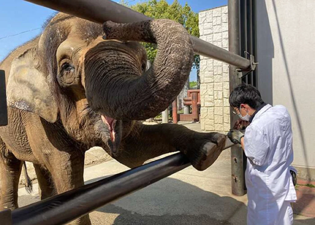
Tuberculosis in Asian Elephants: Interpreting Screening Results in Context
Tuberculosis surveillance in elephants presents a fundamental diagnostic challenge: a test result is...
Article
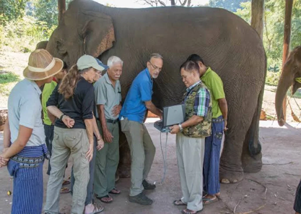
Pregnancy Monitoring in Asian Elephants: Interpreting Ultrasound and Hormonal Findings
Confirming pregnancy is only the beginning of reproductive monitoring in an elephant. With a...