Article
Allergic Otitis Externa in Dogs and Cats: Diagnosis and Management
Allergic otitis externa (AOE) is a frequent manifestation of allergy in companion animals and, in some cases, may be the only clinical sign. It is an inflammatory condition of the ear and should not be confused with primary infection, which often occurs secondarily. While managing secondary infections is important, identifying and addressing the underlying cause of otitis externa is critical to preventing recurrence. According to the 2023 AAHA Management of Allergic Skin Diseases in Dogs and Cats Guidelines1:
- Over half of dogs and around 20% of cats with allergies develop AOE.
- Other primary causes—including parasites, foreign bodies, neoplasia, endocrinopathy, and keratinization disorders—should always be ruled out.
- AOE is frequently associated with atopic dermatitis and food allergy, but not with flea allergy dermatitis.
Clinical Presentation
AOE typically presents as bilateral ear disease, though severity may differ between ears. Key clinical features include:
- Otic pruritus: head shaking, scratching at ears, rubbing face/head
- Pain and erythema
- Ceruminous (waxy) exudate
- Periauricular alopecia and excoriation
Secondary infection may add:
- Malodorous exudate
- Crusting, scaling, and erosions
- Asymmetric ear carriage
Red flags suggesting otitis media/interna:
- Hearing loss
- Head tilt or vestibular disturbances
- Horner’s syndrome
- Temporomandibular joint pain
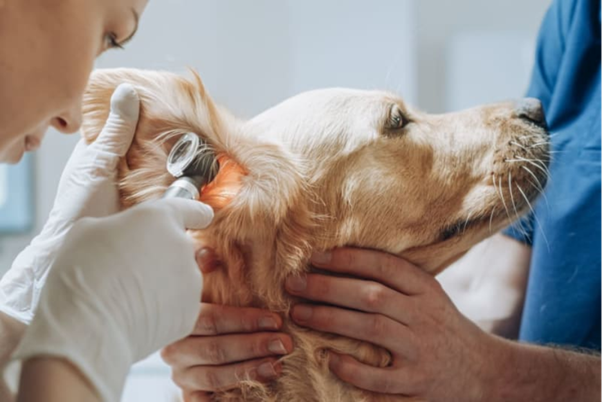
Diagnosis
A thorough ear evaluation is crucial in patients with dermatologic disease, including:
- Examination of pinna and periauricular region
- Palpation of ear canals
- Otoscopy for deeper structures and to rule out other primary causes
Chronic inflammation can cause:
- Altered pliability of ear cartilage, sometimes leading to calcification
- Ceruminous gland hyperplasia → cobblestone-like appearance
- Stenosis of ear canals due to edema, inflammation, or chronic pathologic changes
Secondary infection considerations:
- Dysbiosis is common in dogs with atopic dermatitis; Staphylococcus spp. may predominate
- Yeast overgrowth (Malassezia spp.) is often secondary to inflammatory changes
- Ear cytology is essential in every case:
- Stained dry mount: assesses bacteria and yeast
- Unstained mineral oil wet mount: rules out ectoparasites (important in cats due to Otodectes)
- Training technicians in cytology improves clinic efficiency
Treatment
Topical therapy should be guided by cytologic findings. Key points:
- In cases of abundant exudate, perform in-clinic ear flush
- Be cautious with ruptured tympanic membranes—select safe antimicrobials and cleaning agents
- Referral to a board-certified dermatologist is recommended if:
- Multiple treatment protocols fail
- Disease persists beyond 3–6 months
- Chronic infection complicates management
Short-Term Management
Goals: reduce inflammation and treat secondary infections. Options include:
- Oral corticosteroids (short tapering course)
- Oclacitinib
- Topical corticosteroids for inflammation control
Long-Term Management
Goals: maintain ear health and manage underlying allergy. Strategies include:
- Routine topical maintenance therapy: ear cleansing based on patient needs
- Use of cleansing agents that promote epidermal barrier function and reduce microbial adherence
- Topical glucocorticoids (e.g., hydrocortisone, dexamethasone) to prevent flares and secondary infection
- Monitoring individual response to medications used for atopic dermatitis, as efficacy may vary
Referral indications:
- Complicated or resistant AOE
- Secondary infections unresponsive to empiric therapy
- Middle ear involvement
Owner compliance and follow-up:
- Demonstrate ear cleaning and topical medication application—responsibility of trained technicians
- Interim follow-up via phone, video, or email can improve adherence and outcomes
Conclusion
Allergic otitis externa is a common, often bilateral, manifestation of allergy in dogs and cats that requires careful differentiation from primary infection. Early diagnosis through thorough examination and cytology, combined with targeted topical and systemic therapy, is essential. Long-term management focuses on controlling the underlying allergy, maintaining ear health, and preventing recurrence. Owner education and technician-led follow-up significantly improve treatment success, while referral to a specialist is warranted in complicated or refractory cases.
Reference
- Miller J, Simpson A, Bloom P, Diesel A, Friedeck A, Paterson T, Wisecup M, Yu CM. 2023 AAHA management of allergic skin diseases in dogs and cats guidelines. Journal of the American Animal Hospital Association. 2023 Nov 1;59(6):255-84. https://www.aaha.org/wp-content/uploads/globalassets/02-guidelines/2023-aaha-management-of-allergic-skin-diseases-in-dogs-and-cats-guidelines/resources/2023-aaha-management-of-allergic-skin-diseases-guidelines.pdf.
Related Contents

Upcoming Event
CRIs in Anaesthesia Made Easy
Constant Rate Infusions (CRIs) are widely used in veterinary anaesthesia to provide consistent analg...

Upcoming Event
Homeopathy in Pet Animal Practice
Homeopathy continues to be used by some veterinarians and pet owners as a complementary approach in...
.jpg)
Upcoming Event
Advanced Veterinary Transfusion Medicine
Transfusion medicine has become an essential component of modern veterinary critical care and intern...

Article
PRP, IRAP or Stem Cells? Choosing the Right Biologic for Equine Osteoarthritis
Biologics are everywhere—but which one to choose? Regenerative...

Article
Beyond Wear and Tear: Understanding How Osteoarthritis Develops in Performance Horses
For equine athletes, peak performance and joint health exist in a delicate balance. Whether it is a...

Article
Diagnosing Equine Osteoarthritis Earlier: What Every Equine Vet Should Be Using Today
By the time obvious radiographic changes appear, osteoarthritis (OA) has often been active within th...

Article
Beyond NSAIDs: Practical Treatment Strategies for Equine Osteoarthritis
Managing osteoarthritis (OA) in horses is rarely about finding a single "best" treatment....

Article
The Navicular Case That Won't Improve: What Might Be Getting Missed?
For decades, management of podotrochlear syndrome has focused largely on control...