Article
Practical Diagnosis of Gastric Ulcers in Pigs
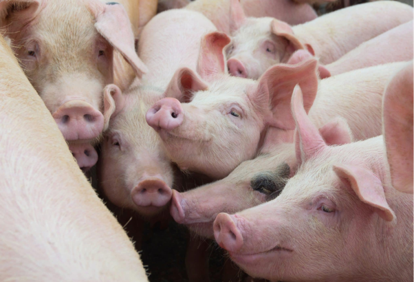
Gastric ulcers are frequently encountered in pigs, yet many affected animals show few or no obvious clinical signs until lesions become severe. As a result, diagnosis often depends on combining clinical observations with post-mortem findings and targeted examination of the stomach. Since lesions are typically confined to the pars esophagea, careful evaluation of this region is essential for accurate diagnosis. A systematic diagnostic approach helps veterinarians assess disease severity, identify herd-level problems, and support management decisions aimed at reducing production losses.
Recognising Clinical Suspicion
Clinical presentation varies according to the severity and duration of ulceration. Peracute and acute cases may result in sudden death due to severe gastric haemorrhage, often without preceding clinical signs1. In contrast, pigs with chronic ulceration may present with less specific signs that can easily be overlooked during routine herd examinations.
Clinical findings that should raise suspicion include:
- Reduced feed intake and growth performance
- Pallor of the eye, oral, or vulvar mucous membranes
- Weakness and reluctance to walk
- Recumbency
- Anorexia
- Melena
- Anaemia characterised by reduced haematocrit, haemoglobin, and erythrocyte count
- Teeth grinding associated with gastric pain
- Greenish vomiting caused by bile staining1
It is important to recognise that pigs with extensive erosive lesions may continue to appear clinically normal when blood loss is minimal1. Therefore, the absence of obvious clinical signs does not exclude significant gastric disease.
Post-Mortem Examination
Necropsy remains an important method for confirming gastric ulceration. Lesions are generally confined to the pars esophagea surrounding the oesophageal opening1.
A healthy pars esophagea appears smooth and white. Early lesions are characterised by thickening, elevation of the mucosa, and yellowish discoloration caused by exposure to hydrochloric acid and bile. Continued irritation leads to progressive erosion and ulcer formation, producing crater-like lesions that may involve large blood vessels and result in severe haemorrhage1.
Ulcers vary considerably in size, depth, and number. Large ulcers often have smooth, raised edges, and extensive lesions may involve most or all of the pars esophagea. Healed lesions frequently leave puckered or star-shaped scars, while severe scarring may narrow the oesophageal opening because of stenosis1.
Importantly, the absence of clotted blood within the stomach should not be interpreted as evidence against gastric ulceration because bleeding may be intermittent1.
Endoscopy and Slaughterhouse Evaluation
Endoscopic examination provides an opportunity to assess the pars esophagea in live pigs. This technique allows direct visualization of erosions and even small bleeding vessels1. It is relatively straightforward to perform once mastered; however, several practical limitations should be considered.
Successful examination requires an empty stomach and adequate restraint under anaesthesia. Feed withdrawal before endoscopy may increase movement of gastric acid and bile into the proximal stomach, potentially increasing the likelihood of ulcer formation during the monitoring period.
Examination of stomachs at slaughterhouses provides valuable herd-level information and remains an important diagnostic tool for identifying macroscopic lesions. After opening and cleaning the stomach, the pars esophagea can be visually assessed and classified according to lesion severity1.
Two scoring systems are described for practical evaluation. One system grades lesions from 0 to 4, ranging from healthy gastric mucosa to ulcers affecting more than 66% of the pars esophagea1,2. Another categorizes lesions as 0 (healthy mucosa), 1 (keratosis), 2 (erosion or mild ulcer), and 3 (severe ulcer)1.
Practical Clinical Insights
Accurate diagnosis depends on evaluating both individual animals and herd-level patterns.
Practical considerations include:
- Include gastric ulceration among differential diagnoses for unexplained anaemia, melena, poor growth, or sudden death.
- Carefully inspect the pars esophagea during every necropsy, even when gastric haemorrhage is not immediately evident.
- Use slaughterhouse findings to monitor lesion prevalence and identify herd-level trends over time.
- Apply lesion scoring systems consistently to support objective assessment and comparison between groups.
- Recognise the strengths and limitations of endoscopy when selecting diagnostic methods for live animals.
A structured diagnostic approach combining clinical assessment, post-mortem examination, endoscopy when appropriate, and slaughterhouse surveillance enables veterinarians to identify gastric ulceration more reliably and supports informed herd health management decisions.
References
- Krepelková Z, Novotný J, Bárdová K, Link R, Csörgö A. Gastric ulcers in pigs–a review. Folia veterinaria. 2024 Jun 27;68(2):33-42. https://sciendo.com/pdf/10.2478/fv-2024-0015
- Marchini CF, Martins PM, Rabelo RN. Prevalence of gastric lesion in pigs. Investigação. 2017;16(8):50-5. https://www.academia.edu/download/125964557/963.pdf
Related Contents

Article
The Elephant is Walking Differently: Where Should the Investigation Begin?
When an elephant begins walking differently, the first challenge is not necessarily to identify the...

Article
When an Elephant’s Blood Report Says “Abnormal”: How Should It Be Interpreted?
A laboratory report can flag a value as abnormal even when an elephant appears clinically healthy. C...
.jpg)
Upcoming Event
World Animal Day
World Animal Day highlights the importance of protecting animal health and welfare through responsib...
Video
Rabies: Epitome of the One Health Paradigm
Rabies exemplifies the One Health approach, linking animal health, human health, veterinary services...
Video
One Health Webinar on Important Updates on Rabies for Veterinary Practitioners and Students
Rabies remains a major zoonotic disease requiring vigilance across veterinary practice and public he...
.jpg)
Video
Transjugular Removal of Canine Heartworms: A Clinical and Surgical Overview
Transjugular removal of canine heartworms is a specialized intervention used in selected dogs with s...

Article
Feline Pregnancy Endocrinology: Hormonal Changes and Placental Function
Pregnancy in cats involves a distinctive endocrine pattern in which ovarian and placental hormones w...

Article
Hormonal Control of Pregnancy Termination in Cats
Pregnancy maintenance in cats depends strongly on coordinated hormonal support, particularly progest...