Article
Enhancing Detection of Canine Testicular Tumors: Role of B-Flow and Contrast-Enhanced Ultrasound
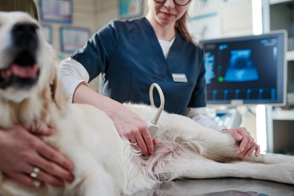
The evaluation of testicular tumors in dogs has traditionally relied on conventional ultrasonography and Doppler-based techniques. While these modalities provide valuable structural and functional information, their limitations in sensitivity and specificity necessitate the use of more advanced imaging approaches. Conventional B-mode ultrasonography, although effective in detecting lesions, cannot reliably characterize tumor types due to highly variable imaging features1,2. Similarly, Doppler techniques may fail to detect vascularization in lesions with small vessels or low blood flow, limiting their diagnostic performance.
These challenges highlight the need for imaging modalities that can better visualize microvascular architecture and improve lesion detection, particularly in cases where early diagnosis is critical for clinical decision-making.
B-Flow Imaging: Improved Visualization of Tumor Vascularity
B-flow imaging represents an important advancement in ultrasound technology. It is based on a digitally encoded technique that suppresses background noise and enhances weak blood flow signals, allowing real-time visualization of intravascular flow using grayscale imaging1. Unlike Doppler methods, B-flow does not rely on frequency shifts, making it less dependent on technical factors such as insonation angle.
This technique demonstrated high sensitivity in detecting vascularization, successfully identifying blood flow patterns in all examined testicular lesions. It also enabled visualization of small vessels that are often not detectable using color Doppler. The vascular distribution patterns observed with B-flow were consistent with those identified using Doppler techniques. Leydig cell tumors showed a predominant perilesional or combined peri/intralesional vascularization pattern, while seminomas, Sertoliomas, and mixed cell tumors were mainly characterized by intralesional vascularization1.
Despite its superior sensitivity, B-flow did not provide sufficient specificity for differentiating tumor types. The overlap in vascular patterns across different tumors limits its role in definitive diagnosis. Additionally, the distribution of arterial sizes did not vary significantly between tumor types, further reducing its discriminatory value.
Contrast-Enhanced Ultrasound: Maximizing Lesion Detection
Contrast-enhanced ultrasound (CEUS) has emerged as a highly sensitive imaging modality for evaluating microvascular perfusion. By using contrast agents, CEUS enhances the visualization of blood flow within tissues, allowing detection of lesions that may not be visible with other techniques.
CEUS successfully identified all testicular tumors, including lesions that were not detected by conventional ultrasonography, Doppler imaging, or B-flow. Notably, a Sertolioma that was missed by other modalities was detected using CEUS, emphasizing its superior sensitivity. No adverse reactions were observed following contrast administration, supporting its safety profile in clinical use1,3,4.
Independent of tumor type, contrast enhancement patterns within lesions differed significantly from those of normal testicular parenchyma. This indicates that CEUS is highly effective in distinguishing abnormal tissue from surrounding normal structures.
Quantitative and Qualitative Assessment with CEUS1
CEUS allows both qualitative and quantitative evaluation of tumor perfusion. Quantitative parameters such as peak intensity (PI), time to peak (TtP), and area under the curve (AUC) were analyzed to assess vascular behavior within lesions.
In certain tumor types, particularly Leydigomas and seminomas, these parameters showed significant differences when compared to normal testicular tissue. However, Sertoliomas and mixed cell tumors did not demonstrate similar variations. Importantly, these quantitative parameters did not differ significantly between tumor types, limiting their usefulness in establishing a definitive diagnosis.
Qualitative assessment of contrast distribution also failed to provide consistent distinguishing features among different tumors. Although enhancement patterns varied between lesions and normal tissue, they were not specific enough to allow reliable tumor classification.
Detection Versus Differentiation: A Clinical Perspective1
A key distinction emerging from these findings is the difference between detection and differentiation. Advanced imaging techniques such as B-flow and CEUS significantly improve the ability to detect testicular tumors, including small or otherwise undetectable lesions. However, their ability to differentiate between tumor types remains limited.
While B-flow enhances visualization of vascular structures and CEUS improves detection of microvascular perfusion, neither modality provides definitive information regarding tumor etiology. The vascular patterns observed across different tumor types show considerable overlap, reducing their specificity.
Conclusion
B-flow and CEUS represent important advancements in the ultrasonographic evaluation of canine testicular tumors. Their ability to enhance visualization of vascularization and detect lesions with greater sensitivity addresses key limitations of conventional and Doppler imaging techniques. However, despite these advantages, their role in differentiating tumor types remains restricted. A multiparametric approach combining structural and functional imaging provides the most comprehensive assessment, improving diagnostic confidence while acknowledging the continued need for further characterization of tumor nature.
Reference
- Orlandi R, Vallesi E, Boiti C, Polisca A, Bargellini P, Troisi A. Characterization of testicular tumor lesions in dogs by different ultrasound techniques. Animals. 2022 Jan 17;12(2):210. https://doi.org/10.3390/ani12020210
- de Souza MB, da Silva LD, Moxon R, Russo M, England GC. Ultrasonography of the prostate gland and testes in dogs. In Practice. 2017 Jan;39(1):21-32. https://www.academia.edu/download/98219257/2017-VETMED.pdf
- Orlandi R, Vallesi E, Boiti C, Polisca A, Troisi A, Righi C, Bargellini P. Contrast-enhanced ultrasonography of maternal and fetal blood flows in pregnant bitches. Theriogenology. 2019 Feb 1;125:129-34. https://www.academia.edu/download/101705762/j.theriogenology.2018.10.02720230501-1-sh77eb.pdf
- Quartuccio M, Mangano C, Macri F, Rizzo M, Di Pietro S, Pugliese M, Mazzullo G, Cristarella S, De Majo M. Contrast-enhanced ultrasound evaluation of testicular interstitial cell tumours in conscious non-sedated dogs. Veterinární medicína. 2018 Mar 1;63(3). https://www.academia.edu/download/98219257/2017-VETMED.pdf
Related Contents

Upcoming Event
CRIs in Anaesthesia Made Easy
Constant Rate Infusions (CRIs) are widely used in veterinary anaesthesia to provide consistent analg...

Upcoming Event
Homeopathy in Pet Animal Practice
Homeopathy continues to be used by some veterinarians and pet owners as a complementary approach in...
.jpg)
Upcoming Event
Advanced Veterinary Transfusion Medicine
Transfusion medicine has become an essential component of modern veterinary critical care and intern...

Article
PRP, IRAP or Stem Cells? Choosing the Right Biologic for Equine Osteoarthritis
Biologics are everywhere—but which one to choose? Regenerative...

Article
Beyond Wear and Tear: Understanding How Osteoarthritis Develops in Performance Horses
For equine athletes, peak performance and joint health exist in a delicate balance. Whether it is a...

Article
Diagnosing Equine Osteoarthritis Earlier: What Every Equine Vet Should Be Using Today
By the time obvious radiographic changes appear, osteoarthritis (OA) has often been active within th...

Article
Beyond NSAIDs: Practical Treatment Strategies for Equine Osteoarthritis
Managing osteoarthritis (OA) in horses is rarely about finding a single "best" treatment....

Article
The Navicular Case That Won't Improve: What Might Be Getting Missed?
For decades, management of podotrochlear syndrome has focused largely on control...