Article
The Role of Computed Tomography in Equine Enterolithiasis: Insights from a Whole-Body CT Case Study
Advances in equine diagnostic imaging continue to expand the clinician's ability to diagnose complex gastrointestinal disorders. A notable example is the application of whole-body computed tomography (CT) in horses with enterolithiasis, providing detailed information that conventional radiography cannot always deliver.
Why CT Matters in Enterolithiasis
Traditional abdominal radiography can often detect enteroliths, but it may not accurately determine their exact size, number, or relationship to surrounding intestinal structures. This limitation becomes particularly important when surgical intervention is planned1.
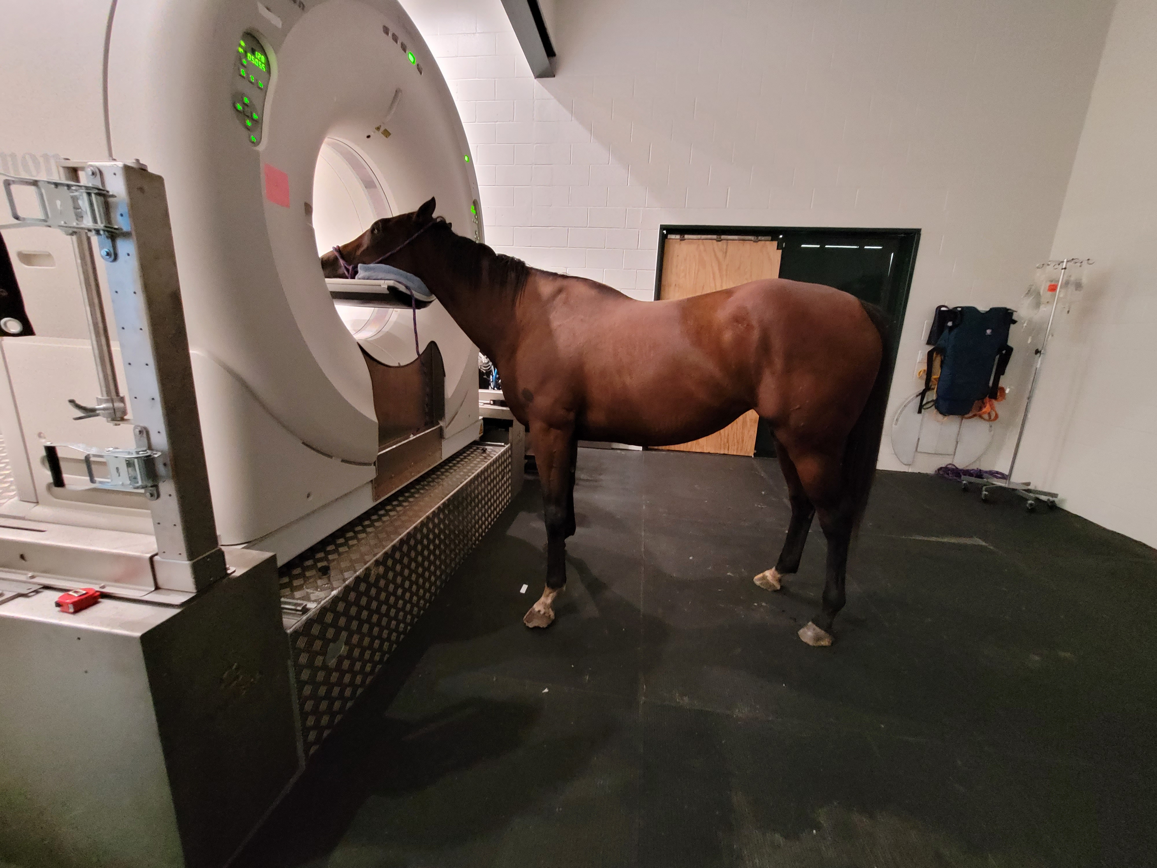
The development of large-gantry CT systems has made it possible to perform whole-body imaging in selected equine patients, offering a new level of diagnostic precision.
A Clinical Case: An 18-Year-Old Pony
An 18-year-old pony presented with chronic anorexia, poor body condition, and weight loss. Initial abdominal radiographs identified a dense oval structure within the abdomen that was consistent with an enterolith1.
To obtain more detailed information, clinicians performed whole-body CT under general anesthesia using a large-bore multidetector scanner. Imaging was acquired with the horse in both prone and supine positions.
What CT Revealed
The CT examination provided several important findings that influenced surgical planning.
Accurate Localization
The enterolith measured approximately 100 mm and was located within the large dorsal colon. Positional imaging demonstrated that the weight of the enterolith caused displacement of the colon within the abdominal cavity, changing its orientation depending on whether the horse was positioned prone or supine.
Identification of Intestinal Displacement
Changing patient position allowed clinicians to visualize gravity-related movement of the gastrointestinal tract and better understand the true anatomical relationships before surgery.
Detection of a Foreign Body Core
One of the most striking findings was the identification of a high-density structure at the center of the enterolith. CT suggested the presence of a metallic foreign body, later confirmed after surgical removal. Three-dimensional reconstruction revealed a structure resembling a staple embedded within the stone.
Surgical Benefits of Preoperative CT
The information obtained from CT significantly improved surgical preparation1,2.
Better Understanding of Anatomy
Knowing the precise location of the affected colon segment reduced exploration time during surgery and allowed surgeons to anticipate anatomical displacement before entering the abdomen.
Accurate Assessment of Size
CT provided precise measurements of the enterolith, helping surgeons determine whether manipulation through the intestinal tract would be possible or whether enterotomy would be required.
Identification of Multiple Enteroliths
Approximately 40% of horses with enterolithiasis may harbor multiple enteroliths1. CT enables clinicians to evaluate the entire abdomen and identify additional stones that may be missed with conventional radiography.
Surgical Outcome1
During celiotomy, surgeons confirmed the CT findings and removed a 104 mm enterolith weighing 422 g from the large dorsal colon. Mineral analysis demonstrated a composition of approximately 50% magnesium ammonium phosphate and 50% calcium phosphate.
The horse recovered uneventfully, regained appetite, and increased body weight from 158 kg to 205 kg within six months after surgery.
Future Applications of Whole-Body CT
This case highlights the growing value of advanced imaging in equine gastrointestinal surgery. Whole-body CT not only improves identification of enterolith location, size, and number but also provides critical information regarding intestinal displacement and potential foreign-body niduses.
As access to large-bore CT systems increases, this technology may become an increasingly valuable adjunct for the diagnosis and surgical management of complex equine colic cases involving enterolithiasis.
References
- Nakamae Y, Ishihara A, Itoh M, Yanagawa M, Sasaki N, Yamada K. Displacement of the large colon in a horse with enterolithiasis due to changed positions observed by computed tomography. Journal of Equine Science. 2018;29(1):9-13. https://www.jstage.jst.go.jp/article/jes/29/1/29_1728/_pdf
- Taylor CJ, Peter VG, Coleridge MO, Bathe AP. Immediate pre-operative computed tomography for surgical planning of equine fracture repair: A retrospective review of 55 cases. Plos one. 2022 Dec 28;17(12):e0278748. https://journals.plos.org/plosone/article/file?id=10.1371/journal.pone.0278748&type=printable
Related Contents

Upcoming Event
African Swine Fever: Diagnosis and Management
African swine fever can cause severe disease and significant losses in affected pig populations.&nbs...
.jpg)
Video
Transjugular Removal of Canine Heartworms: A Clinical and Surgical Overview
Transjugular removal of canine heartworms is a specialized intervention used in selected dogs with s...

Article
Feline Pregnancy Endocrinology: Hormonal Changes and Placental Function
Pregnancy in cats involves a distinctive endocrine pattern in which ovarian and placental hormones w...

Article
Hormonal Control of Pregnancy Termination in Cats
Pregnancy maintenance in cats depends strongly on coordinated hormonal support, particularly progest...

Article
Feline Embryo Implantation and Early Placental Development
Pregnancy establishment in cats involves a sequence of precisely timed events between fertilization,...

Article
Ultrasonographic Monitoring of Pregnancy in Cats: A Gestational Timeline
Ultrasonography provides a practical way to follow the progression of feline pregnancy by identifyin...

Article
Feline Mismating: Pregnancy Diagnosis and Clinical Management
Mismating or unplanned mating is a common clinical problem in sexually mature queen cats because of...

Article
Medical Termination of Pregnancy in Queens: Drugs, Timing and Treatment Protocols
Medical termination of pregnancy (MTP) in queen cats should be approached according to the timing of...