Article
Diagnosing Tuberculosis in Camels: A Practical Diagnostic Approach
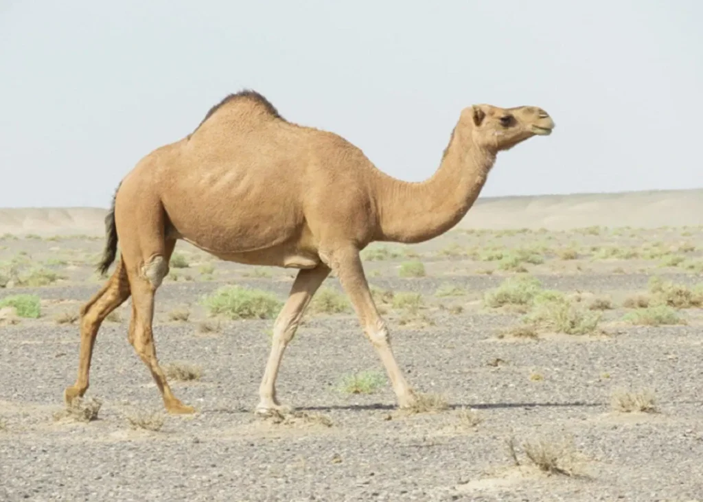
A camel may appear clinically normal while harbouring advanced tuberculous lesions. That makes diagnosis particularly challenging: should suspicion rely on clinical signs alone, or should veterinarians look beyond the obvious?
Tuberculosis in camels is a chronic disease, and many infected animals remain asymptomatic until the disease is advanced. Clinical signs can include wasting, anorexia, weakness, chronic weight loss or emaciation, cough, dyspnea, respiratory distress, and enlargement of superficial lymph nodes. However, severe pulmonary lesions may sometimes occur without obvious respiratory distress, and animals may occasionally be found dead without previous clinical observations[1].
Start with the clinical examination, but do not stop there[1]
Physical examination should include body temperature, pulse rate, respiratory rate, nasal discharge when present, regional lymph nodes, and visible mucous membranes.
The challenge is that these findings are not sufficient to confirm tuberculosis. Because the disease can involve different organs and produce variable clinical signs, tuberculosis may need to remain in the differential diagnosis when chronic or unexplained disease is present.
Post-mortem examination can reveal the pattern[1]
In suspected cases, systematic examination of the lymph nodes and organs is important. Mandibular, retropharyngeal, bronchial, mediastinal, mesenteric, and hepatic lymph nodes should be examined, along with the lungs, liver, small intestine, and kidneys. The lungs are inspected and palpated externally, then sectioned to facilitate detection of lesions; lymph nodes are similarly sliced and examined for visible abnormalities.
Typical lesions include yellowish, caseous nodules with friable centres. They may occur in the lungs, liver, spleen, and several lymph nodes. Cut sections can show firm, yellowish nodules with an onionskin-like structure and partially mineralised centres.
What can tuberculin testing tell you?[1],[2]
Two skin tests are described for camel tuberculosis: The single intradermal tuberculin test (SITT) and the single comparative intradermal tuberculin test (CITT).
For SITT, bovine PPD is administered and changes in skin thickness are recorded on days 2, 3, 5 and 7. A difference of <2.0 mm is considered negative, 2.0–4.0 mm suspicious and >4.0 mm a reactor (Wernery et al., 2007).
CITT compares reactions to bovine and avian PPD. A bovine reaction >4.0 mm greater than the avian reaction is considered positive, while a 1.0–4.0 mm difference is inconclusive.
Yet ante-mortem diagnosis remains difficult because adequate tests for live camelids are limited.
Confirmation requires more than one finding[1]
Definitive diagnosis can be established at post-mortem through characteristic lesions followed by histopathology and bacterial culture. Ziehl-Neelsen staining can demonstrate acid-fast bacilli, while culture remains a key confirmatory method, although mycobacteria grow slowly and may require prolonged incubation on selective media such as Lowenstein-Jensen or Ogawa.
PCR provides another diagnostic option and can detect very low levels of Mycobacterium tuberculosis complex DNA. However, reported sensitivity varies, particularly between smear-positive and smear-negative samples, and amplification methods should not currently replace conventional culture.
Clinical bottom line: In camel tuberculosis, a normal-looking animal does not reliably exclude infection. Clinical assessment, tuberculin testing, pathological examination and laboratory confirmation each provide different pieces of the diagnostic picture, making a combined approach particularly important.
References:
- Yessuf JA, Jibril Y, Tekle Y, Mammo G. Review on camel tuberculosis: diagnosis, pathology and public health significance. World J Pharm Life Sci. 2016. https://www.researchgate.net/publication/361485849
- Wernery U, Kinne J, Jahans KL, Vordermeier HM, Esfandiari J, Greenwald R, Johnson B, Ul-Haq A, Lyashchenko KP. Tuberculosis outbreak in a dromedary racing herd and rapid serological detection of infected camels. Veterinary microbiology. 2007 May 16;122(1-2):108-15. https://www.sciencedirect.com/science/article/abs/pii/S0378113507000375
Related Contents
_Resize_2.jpg)
Upcoming Event
Leptospirosis in Animals: Diagnosis and Zoonotic Prevention
Leptospirosis can cause significant illness in animals and pose a zoonotic risk to humans. Explore c...
Article
Sudden Death in Cattle: A Practical Differential Diagnosis
A cow is found dead during the morning inspection. There were no obvious warning signs the previous...
Article
Caudal Vena Cava Thrombosis in Cattle: Recognising a Fatal Complication of Liver Abscessation
A cow may appear completely healthy until sudden respiratory distress, haemoptysis or death occurs....
Article
Sudden Death in Sheep: Key Differential Diagnoses for the Veterinarian
A sheep found dead on pasture may leave little warning. One animal may have been affected by a rapid...
Article
Sudden Death in Goats: Recognising Peracute Infectious and Metabolic Causes
A goat found dead with little or no warning can leave very few clinical clues. In these cases, the p...
Article
Necrotic Enteritis in Broilers: Identifying the Farm-Level Risk Factors
A broiler flock showing wet litter, poor performance or rising mortality may have several problems o...
Article
Necrotic Enteritis in Broilers: Linking Intestinal Lesions with Laboratory Diagnosis
A broiler may arrive for postmortem examination with a distended, fragile small intestine covered by...
Article
Controlling Necrotic Enteritis Without Antibiotic Growth Promoters: What Can Feed-Based Strategies Offer?
When antibiotic growth promoters are no longer the main option for controlling necrotic enteritis (N...