Article
Managing Ovine Footrot in Clinical Practice: Lessons from Successful Clinical Management
Footrot remains one of the most important causes of lameness and economic loss in sheep production. The condition not only compromises animal welfare but also affects flock productivity through reduced weight gain, decreased reproductive performance, increased treatment costs, and premature culling1,2,3. Since multiple environmental and management factors influence disease occurrence, successful clinical management requires more than simply treating the affected hoof. Understanding the clinical presentation, recognizing predisposing factors, and implementing appropriate wound care and antimicrobial therapy can significantly improve outcomes1.
Recognizing the Clinical Picture
The severity of ovine footrot can vary depending on the virulence of the causative organisms, environmental conditions, trauma to the interdigital skin, and host-related factors1. Lameness is often the most noticeable clinical sign, although animals with early infection may not initially show obvious gait abnormalities2.
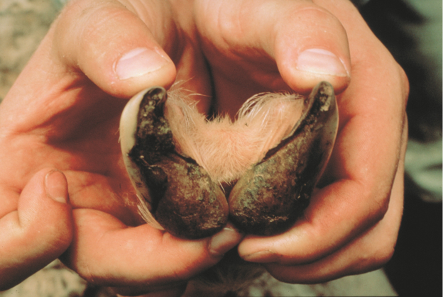
An adult Abergele sheep presented with non-weight-bearing lameness of the left hindlimb following progressive deterioration of the hoof. Clinical examination revealed depression, reduced appetite, swelling of the interdigital space, heel erosion, accumulation of necrotic tissue, dirt, and foul-smelling purulent discharge. The hoof had split, and the interdigital region contained debris, producing the characteristic offensive odor associated with footrot. Elevated physiological parameters, including rectal temperature, heart rate, and respiratory rate, further supported the presence of an active inflammatory process.
Looking Beyond the Hoof
Environmental assessment can provide valuable clues during clinical evaluation. In this case, the sheep originated from a poorly maintained intensive production system where pens were cleaned only once weekly. The disease occurred during the wet season, and the affected environment received substantial annual rainfall, conditions known to favor disease transmission1,2.
The owner also reported similar hoof problems among other sheep within the flock and in nearby farms, emphasizing the contagious nature of the condition and the importance of evaluating flock-level risk alongside individual animal management.
Footrot should also be differentiated from other causes of lameness, including hoof separation, white line lesions, foot abscesses, sole ulceration, foreign body injuries, granulomatous lesions, severely overgrown feet, treponeme infections, laminitis, and primary claw horn lesions with secondary infection1,4. Careful clinical examination, supported by laboratory diagnostics where indicated, assists in establishing an accurate diagnosis1,2.
Practical Approach to Clinical Management1
Successful management combined meticulous local wound care with systemic therapy.
The affected hoof was:
- Thoroughly cleaned using isotonic saline solution
- Disinfected with 3% hydrogen peroxide followed by 1% povidone-iodine
- Treated with oxytetracycline (10 mg/kg, intramuscularly) for five consecutive days
- Managed with meloxicam (0.5 mg/kg, intramuscularly) three times daily for three days to reduce pain and inflammation
- Treated locally using topical oxytetracycline spray following cleaning and during subsequent dressing sessions
Follow-up care included repeated dressing with 1% povidone-iodine on the second and third days after treatment, with continued monitoring until the wound dried and healthy healing was evident. Within seven days, the hoof showed marked improvement, and four weeks later, the sheep had regained good overall health and welfare. Along with treatment, the owner was advised to improve pen hygiene, provide quality feed, and clean the hooves regularly to minimize contamination and support recovery.
Key Takeaways
- Early recognition of characteristic hoof lesions and lameness is essential for timely intervention.
- Combining thorough hoof cleaning, disinfection, systemic therapy, and topical treatment may improve clinical outcomes.
- Good housing hygiene and regular hoof care remain important components of footrot management and may help reduce disease spread within the flock.
References
- Fesseha H. Ovine footrot and its clinical management. Veterinary Medicine: Research and Reports. 2021 May 11:95-9. https://www.tandfonline.com/doi/pdf/10.2147/VMRR.S306207
- Groenevelt M, Anzuino K, Smith S, Lee MR, Grogono-Thomas R. A case report of lameness in two dairy goat herds; a suspected combination of nutritional factors concurrent with treponeme infection. BMC research notes. 2015 Dec 16;8(1):791. https://link.springer.com/content/pdf/10.1186/s13104-015-1734-3.pdf
- Winter JR, Kaler J, Ferguson E, KilBride AL, Green LE. Changes in prevalence of, and risk factors for, lameness in random samples of English sheep flocks: 2004–2013. Preventive veterinary medicine. 2015 Nov 1;122(1-2):121-8. https://wrap.warwick.ac.uk/id/eprint/72942/8/WRAP_prevalence%20and%20risk%20July%202015%20final%20full%20paper.pdf
- Bitrus AA, Abba Y, Jesse FF, Yi LM, Teoh R, Sadiq MA, Chung EL, Lila MA, Haron AW. Clinical management of foot rot in goats: A case report of lameness. Journal of Advanced Veterinary and Animal Research. 2017 May 8;4(1):110-6. https://banglajol.info/index.php/JAVAR/article/view/32480/21973
Related Contents
.jpg)
Video
Transjugular Removal of Canine Heartworms: A Clinical and Surgical Overview
Transjugular removal of canine heartworms is a specialized intervention used in selected dogs with s...

Article
Feline Pregnancy Endocrinology: Hormonal Changes and Placental Function
Pregnancy in cats involves a distinctive endocrine pattern in which ovarian and placental hormones w...

Article
Hormonal Control of Pregnancy Termination in Cats
Pregnancy maintenance in cats depends strongly on coordinated hormonal support, particularly progest...

Article
Feline Embryo Implantation and Early Placental Development
Pregnancy establishment in cats involves a sequence of precisely timed events between fertilization,...

Article
Ultrasonographic Monitoring of Pregnancy in Cats: A Gestational Timeline
Ultrasonography provides a practical way to follow the progression of feline pregnancy by identifyin...

Article
Feline Mismating: Pregnancy Diagnosis and Clinical Management
Mismating or unplanned mating is a common clinical problem in sexually mature queen cats because of...

Article
Medical Termination of Pregnancy in Queens: Drugs, Timing and Treatment Protocols
Medical termination of pregnancy (MTP) in queen cats should be approached according to the timing of...

Article
Safety and Aftercare Following Pregnancy Termination in Queen Cats
Pregnancy termination in queen cats requires more than selecting an appropriate abortifacient. Once...