Article
Finding the Cause of Seizures: A Practical Diagnostic Approach
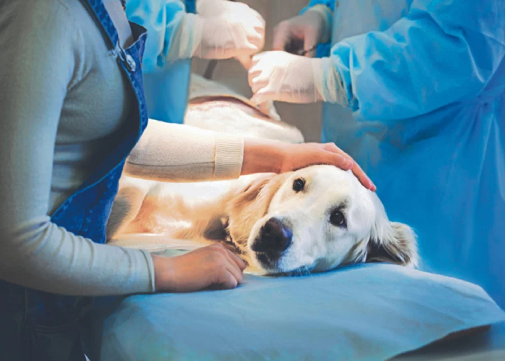
A seizure has stopped, but the diagnostic work is not over. The immediate question is not simply whether the patient has a brain disease. Could a metabolic, toxic, vascular, or other extracranial problem be driving the seizure activity?
For dogs and cats presenting with seizures, the diagnostic approach should begin with potentially reversible extracranial causes before moving toward advanced investigation of intracranial disease.
Start With Rapid Point-of-Care Assessment
Several extracranial causes can be investigated quickly during the initial work-up. Blood collected during intravenous catheter placement can be used to assess:
- Blood glucose
- Packed cell volume
- Total proteins
- Lactate
- Electrolytes with venous blood gas analysis
A systolic blood pressure measurement can also help identify severe hypertension.
Hypoglycemia is one immediately treatable cause. A blood glucose concentration below 3.3 mmol/L is used to identify hypoglycemia[1]. Other metabolic possibilities include uremia, hepatic encephalopathy, hyperlipidemia, sodium or calcium abnormalities, and hyperosmolar disorders.
History remains important alongside these tests. Medication exposure, underlying disease, and possible access to toxins can change the differential substantially. Toxic causes listed in the clinical framework include lead, organophosphates, chlorinated hydrocarbons, xylitol, caffeine, and ethylene glycol[2].
Do Not Stop at Routine Blood Testing[2]
Some suspected causes require a targeted investigation.
When hepatic encephalopathy is a concern, fasted ammonia levels and pre- and post-prandial bile acid testing may be appropriate. Coagulation testing, including PT/aPTT or thromboelastography, and specific infectious disease testing may also be indicated depending on the clinical presentation.
The objective is not to perform every available test indiscriminately. It is to use the history, physical examination, and initial findings to determine which causes require further investigation.
Use Signalment and Clinical Course to Prioritize Differentials[2]
Age can provide an important diagnostic clue.
Congenital and infectious abnormalities are more likely considerations in young animals, whereas neoplastic or degenerative intracranial disease becomes more common in geriatric patients.
The pattern of onset and progression can also help distinguish differentials. An older dog with a primary brain tumour may develop seizures later in life, with neurologic signs becoming progressively worse as the tumour grows, and peri-tumoral inflammation contributes to further structural change. In contrast, a middle-aged dog experiencing an ischemic event may develop more severe signs acutely, followed by improvement over time.
These patterns do not establish a diagnosis on their own, but they can help determine which differentials deserve greater attention.
Organize the Differential With DAMNIT-V
When multiple possibilities remain, the DAMNIT-V mnemonic provides a practical framework for organizing seizure differentials[2]:
- Degenerative
- Anomalous
- Metabolic
- Neoplastic
- Inflammatory/Infectious
- Trauma/Toxin
- Vascular
Intracranial causes within these categories include conditions such as hydrocephalus, storage diseases, meningioma, glioma, lymphoma, infectious or immune-mediated disease, traumatic brain injury, ischemia, and hemorrhage.
Extracranial causes include metabolic abnormalities, toxicities, severe hyperthermia, blood dyscrasias, hypertension, and hyperviscosity.
When is Advanced Imaging Appropriate?
If an extracranial cause has not explained the seizure activity and intracranial disease remains a concern, definitive investigation may require computed tomography, magnetic resonance imaging, or cerebrospinal fluid analysis[2]. These diagnostics may not be readily available in general practice or emergency settings.
That makes the initial clinical assessment especially valuable. A combination of signalment, history, physical examination and readily available diagnostic testing can help determine whether the next step should be correction of an identified systemic abnormality or further investigation of intracranial disease.
Clinical Bottom Line
A seizure should trigger a structured search for its cause, not an immediate assumption of primary brain disease. Rapidly assess reversible extracranial abnormalities, use signalment and clinical progression to prioritize differentials, and apply DAMNIT-V to keep the broader diagnostic framework organized. Advanced diagnostics can then be considered when the clinical picture continues to point toward intracranial disease.
References:
- Idowu O, Heading K. Hypoglycemia in dogs: Causes, management, and diagnosis. The Canadian Veterinary Journal. 2018 Jun;59(6):642. https://pmc.ncbi.nlm.nih.gov/articles/PMC5949948/pdf/cvj_06_642.pdf
- Munguia GG, Brooks AC, Thomovsky SA, Thomovsky EJ, Rincon A, Johnson PA. Emergency approach to acute seizures in dogs and cats. Veterinary Sciences. 2024 Jun 17;11(6):277. https://www.mdpi.com/2306-7381/11/6/277
Related Contents
Article
FGF-23 in Feline CKD: An Early Signal of Phosphate Dysregulation
A cat with early chronic kidney disease (CKD) may have a normal serum phosphate concentration. Does...
Article
Albuminuria or Transferrinuria? Reading the Glomerular Signal in Feline CKD
A cat can have chronic kidney disease (CKD) with only mild proteinuria. So, when urinary protein is...
Article
Tubular Injury Biomarkers in Feline CKD: What Can NGAL, KIM-1 and L-FABP Tell You?
When feline CKD is already suspected, identifying reduced renal function is only part of the clinica...
Article
Can Renal Fibrosis Biomarkers Reveal CKD Progression in Cats?
Chronic kidney disease in cats is not only a problem of declining renal function. Structural changes...
Article
When Creatinine May Not Tell the Full Story in Feline Acute Kidney Injury
Serum creatinine is routinely used with changes in urine output to diagnose and grade Acute Kidney I...
Article
When a Normal-Looking Udder Hides Mastitis: Recognising Subclinical Mastitis in Buffaloes
A buffalo can appear clinically normal while mastitis is already affecting the udder and milk produc...
Article
Buffalo Subclinical Mastitis: Which Risk Factors Should Vets Investigate?
When subclinical mastitis (SCM) keeps appearing in a buffalo herd, looking only at the affected anim...
Article
Diagnosing Subclinical Mastitis in Buffaloes: From SCC and CMT to Molecular Tests
Subclinical mastitis (SCM) can remain hidden because the buffalo may show no obvious udder or milk a...