Article
Canine Pyoderma: A Stepwise Approach to Diagnosis and Sampling
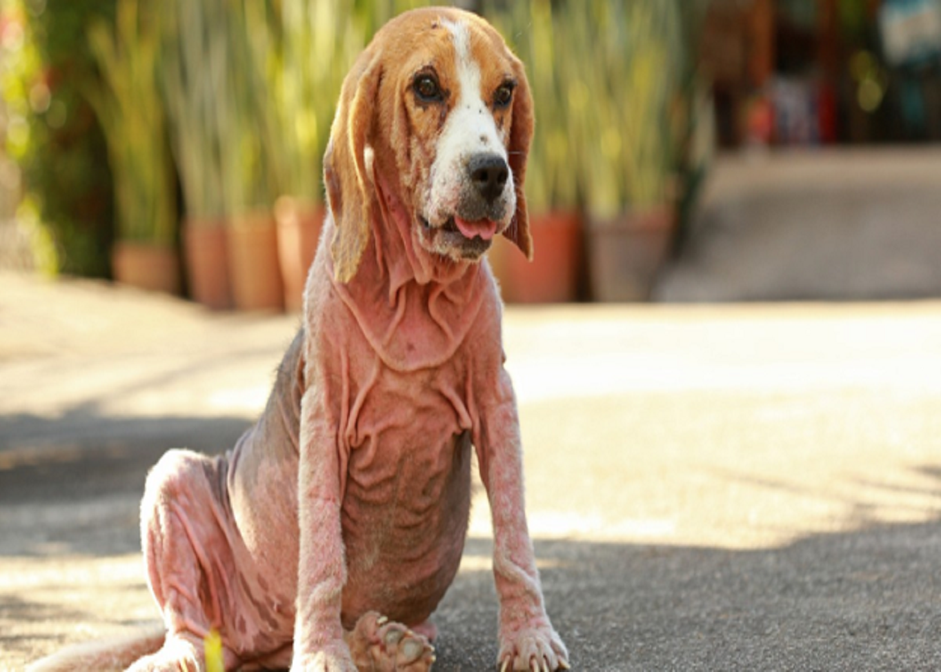
A dog presents with erythema, pustules, crusting or draining skin lesions. Are these findings sufficient to diagnose bacterial pyoderma? Not necessarily. Several non-infectious skin conditions can produce similar clinical signs, making diagnostic confirmation essential before initiating antimicrobial treatment. A systematic approach combining complete skin examination, cytology and investigation of underlying causes can help veterinarians improve diagnostic accuracy and avoid unnecessary antimicrobial use.
Start With a Complete Skin Examination[1]
The first step is to examine the entire skin surface and assess the distribution, extent and type of lesions. Long hair may need to be trimmed around affected areas to improve visibility and facilitate sampling. Scissors are generally preferred over clippers to avoid damaging lesions before diagnostic evaluation.
The depth of infection also influences the diagnostic and treatment approach. Canine pyoderma is classified into three categories:
- Surface pyoderma: Includes pyotraumatic acute moist dermatitis and intertrigo.
- Superficial pyoderma: Commonly presents as superficial bacterial folliculitis, with papules, follicular pustules, crusts and epidermal collarettes.
- Deep pyoderma: May involve furunculosis, cellulitis, ulcers, nodules, fistulae and haemopurulent discharge.
No single lesion is diagnostic of a particular infection depth. Clinical findings must be interpreted together, particularly when superficial and deep presentations overlap.
Why Cytology Should Be Part of Every Skin Examination[1],[2]
Cytology is recommended in every suspected case of canine pyoderma to confirm bacterial involvement. It is a practical in-house investigation that provides rapid results without requiring complicated staining procedures.
The presence of intracellular cocci, extracellular bacteria with nuclear streaming or large numbers of bacteria without inflammatory cells can support a diagnosis of bacterial infection.
Cytology also helps differentiate pyoderma from conditions such as Malassezia dermatitis and pemphigus foliaceus. Its diagnostic sensitivity has been reported as 93% in canine superficial pyoderma.
Importantly, cytology is not limited to initial diagnosis. It can also help monitor treatment response and confirm resolution of infection.
Collecting Representative Samples[1]
Sampling technique should be selected according to lesion type and infection depth.
- Pustules: Aspirate or lance intact pustules using a sterile needle and collect their contents.
- Crusts and epidermal collarettes: Sample the exposed skin beneath the crust or the leading edge of the collarette.
- Dry lesions: Tape preparations or swabs can help collect diagnostic material.
- Deep lesions: Tissue biopsy or fine-needle aspiration may be necessary to obtain representative samples.
For deep pyoderma, surface swabs can be misleading. In dogs with acral lick dermatitis, surface culture predicted deep tissue isolates in only 8 of 22 cases.
Investigate the Underlying Cause[1]
Canine pyoderma is always secondary to an underlying condition or predisposing factor. Atopic dermatitis, ectoparasitic infestations, dermatophytosis, endocrine disorders and cornification abnormalities can contribute to infection.
A minimum diagnostic assessment should include coat brushings, hair plucks and skin scrapings to investigate ectoparasites and dermatophytes. Further testing may be indicated depending on clinical findings.
Clinical Bottom Line
Accurate diagnosis of canine pyoderma depends on more than recognising skin lesions. Complete examination, routine cytology, appropriate sample collection and investigation of underlying causes provide a structured foundation for treatment decisions and responsible antimicrobial stewardship.
References:
- Loeffler A, Cain CL, Ferrer L, Nishifuji K, Varjonen K, Papich MG, Guardabassi L, Frosini SM, Barker EN, Weese JS. Antimicrobial use guidelines for canine pyoderma by the International Society for Companion Animal Infectious Diseases (ISCAID). Veterinary dermatology. 2025 Jun;36(3):234-82. https://onlinelibrary.wiley.com/doi/pdf/10.1111/vde.13342
- Udenberg TJ, Griffin CE, Rosenkrantz WS, Ghubash RM, Angus JC, Polissar NL, Neradilek MB. Reproducibility of a quantitative cutaneous cytological technique. Veterinary dermatology. 2014 Oct;25(5):435-e67. https://onlinelibrary.wiley.com/doi/10.1111/vde.12138
Related Contents
Article
Recognising Leptospirosis Across Animal Species: Clinical Presentations and Organ-Specific Changes
A dog presents with fever and systemic illness, while cattle experience reproductive failure without...
Article
Psychogenic Polydipsia in Dogs: Recognising Behavioural Triggers and Clinical Indicators
A dog presents with persistent excessive drinking and urination, yet its physical examination reveal...
Article
Diagnostic Approach to Canine PU/PD: Differentiating Primary Polydipsia from Primary Polyuria
Persistent excessive drinking and urination can be the presenting signs of several conditions in dog...
.jpg)
Upcoming Event
Culture-Guided Treatment of UTIs in Dogs and Cats
Make informed antibiotic choices in canine and feline UTIs with a culture-guided approach. Explore h...
Article
Managing Psychogenic Polydipsia in Dogs: Water Intake, Behavioural Therapy and Pharmacological Intervention
Once psychogenic polydipsia (PPD) has been identified, management needs to address more than the vol...
Article
Duck Viral Hepatitis in Ducklings: Clinical Recognition and Differential Diagnosis
A sudden rise in mortality among young ducklings, particularly those under three weeks of age, shoul...
Article
Confirming Duck Viral Hepatitis: Choosing and Interpreting Diagnostic Tests
When young ducklings develop sudden mortality and liver lesions suggestive of duck viral hepatitis (...
Article
Duck Viral Hepatitis Vaccination: Managing Maternal Antibody Interference and Field Efficacy
Vaccination is a key strategy for controlling Duck Viral Hepatitis (DVH), particularly in intensive...