Article
Clinical Recognition of Caseous Lymphadenitis in Sheep and Goats
Caseous lymphadenitis (CLA), commonly referred to as "cheese gland," is a chronic bacterial disease of sheep and goats that remains a significant health and economic concern in small ruminant production. The disease is caused by Corynebacterium pseudotuberculosis biovar ovis and is characterized by pyogranulomatous abscess formation in superficial lymph nodes and internal organs. Because infected animals may remain in the flock for prolonged periods while continuously serving as a source of infection, early clinical recognition is essential for effective disease management1,2.
For practicing veterinarians, recognizing the different clinical presentations of CLA is the first step toward timely diagnosis, limiting transmission, and implementing appropriate flock-level interventions.
Understanding the Clinical Forms
CLA presents in two primary forms: external (cutaneous or superficial) and internal (visceral). Both forms may occur independently or coexist in the same animal3.
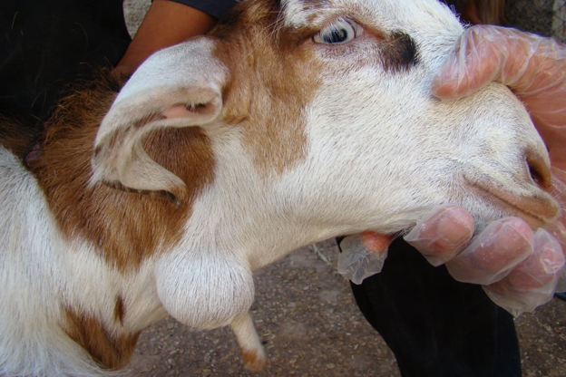
The external form is the one most frequently encountered during routine clinical examinations. It is characterized by abscess formation in superficial lymph nodes, particularly the mandibular, parotid, pre-scapular, superficial cervical, subiliac, popliteal, and supramammary lymph nodes. In some cases, swelling may also develop within surrounding subcutaneous tissues4,5.
The visceral form is more challenging because lesions develop within internal organs such as the lungs, liver, spleen, kidneys, uterus, and internal lymph nodes including the mediastinal, bronchial, and lumbar lymph nodes4,6,7.
What to Look for During Clinical Examination
Superficial CLA abscesses typically appear as firm, well-defined, painless swellings beneath the skin. Over time, these lesions enlarge and become encapsulated, often remaining present for months or even years before rupturing.
Once mature, the abscess usually opens through a fistula and releases a thick, odourless, greenish-white purulent exudate. This material contains viable bacteria capable of contaminating the surrounding environment. Following drainage, the lesion gradually heals with scar formation, although recurrence months or years later is common because the infection is rarely eliminated completely4,6,8.
An important species difference should also be considered during examination. In sheep, abscesses commonly develop an onion-like appearance with concentric fibrous layers surrounding caseous material. In goats, lesions generally contain a dry, homogeneous purulent paste rather than the characteristic layered structure seen in sheep5,8.
Recognizing the Less Obvious Cases
One of the greatest clinical challenges is identifying animals with visceral CLA. Unlike the external form, these animals often exhibit few obvious abnormalities during physical examination.
When clinical signs are present, they may include chronic weight loss, fever, cough, or purulent nasal discharge. Because these signs are non-specific, visceral CLA frequently remains undetected until post-mortem examination4.
In sheep, chronic emaciation associated with visceral infection is often described as thin-ewe syndrome. Unless affected animals develop progressive weight loss and weakness, general appearance alone rarely allows reliable identification of internal disease3.
The distribution of disease may also differ between species. Goats are more likely to develop the external form, whereas sheep more commonly develop visceral lesions. Reports indicate that fewer than 20% of CLA cases in goats involve the internal form9,10.
Practical Clinical Insights
During routine flock visits, veterinarians should maintain a high level of suspicion whenever persistent lymph node enlargement is detected. Particular attention should be given to animals with recurrent abscesses, chronic weight loss, or lesions located around major peripheral lymph nodes.
Because apparently healthy animals may harbour internal infections or develop recurrent abscesses long after previous lesions have healed, clinical examination should always be considered the starting point rather than the endpoint of disease investigation. Careful recognition of characteristic lesions allows earlier intervention and helps reduce ongoing contamination within the flock.
Take-Home Message
Successful management of CLA begins with accurate clinical recognition. Identifying the characteristic superficial abscesses, remaining alert for subtle signs of visceral disease, and understanding the different presentations in sheep and goats enables veterinarians to detect affected animals earlier and make informed decisions regarding further diagnostic testing and flock health management.
References
- Bettini A, Mancin M, Mazzucato M, Schanung A, Colorio S, Tavella A. A seroepidemiological survey of Corynebacterium pseudotuberculosis infection in South Tyrol, Italy. Pathogens. 2022 Nov 9;11(11):1314. https://www.mdpi.com/2076-0817/11/11/1314
- Oreiby AF. Diagnosis of caseous lymphadenitis in sheep and goat. Small Ruminant Research. 2015 Jan 1;123(1):160-6. https://www.academia.edu/download/119926157/j.smallrumres.2014.11.01320241201-1-iv1aar.pdf
- Dorella F, Pacheco LG, Oliveira S, Miyoshi A, Azevedo V. Corynebacterium pseudotuberculosis: microbiology, biochemical properties, pathogenesis and molecular studies of virulence. Veterinary research. 2006;37(2):201-18. https://hal.science/hal-00903023/document
- Dopuđ M, Reil I, Zdelar-Tuk M, Špičić S, Duvnjak S. Caseous Lymphadenitis in sheep and goats–“Cheese Glands”. Veterinarska stanica. 2025;56(3):303-16. https://hrcak.srce.hr/file/462890
- Ruiz H, Ferrer LM, Ramos JJ, Baselga C, Alzuguren O, Tejedor MT, de Miguel R, Lacasta D. The relevance of caseous lymphadenitis as a cause of culling in adult sheep. Animals. 2020 Oct 24;10(11):1962. https://www.mdpi.com/2076-2615/10/11/1962
- de Sá Guimarães A, do Carmo FB, Pauletti RB, Seyffert N, Ribeiro D, Lage AP, Heinemann MB, Miyoshi A, Azevedo V, Guimarães Gouveia AM. Caseous lymphadenitis: epidemiology, diagnosis, and control. IIOAB J. 2011;2(11). https://www.researchgate.net/profile/Alessandro-Guimaraes/publication/285967469
- Thongkwow S, Poosiripinyo N, Pongkornkumpon N, Saengsakchai S, Klinkhiew N, Chalatan T, Kanistanon K, Lerk-u-suke S, Rerkyusuke S. Distribution and risk factors of clinical caseous lymphadenitis in small-holder goat herds in Northeastern Thailand. The Thai Journal of Veterinary Medicine. 2019;49(4):343-51. https://digital.car.chula.ac.th/cgi/viewcontent.cgi?article=2999&context=tjvm
- Habte D. Caseous lymphadenitis: A case of sheep and its management in Ethiopia. Ethiopian Veterinary Journal. 2023 Aug 8;27(2):187-95. https://www.ajol.info/index.php/evj/article/view/252512/238547
- Guimarães AS, Seyffert N, Bastos BL, Portela RW, Meyer R, Carmo FB, Cruz JC, McCulloch JA, Lage AP, Heinemann MB, Miyoshi A. Caseous lymphadenitis in sheep flocks of the state of Minas Gerais, Brazil: prevalence and management surveys. Small Ruminant Research. 2009 Nov 1;87(1-3):86-91. https://www.sciencedirect.com/science/article/pii/S0921448809001850
- Habuš J, Matanović K, Majetić ZŠ, Rukavina T, Ćorić A, Milas Z, Starešina V, Martinec BŠ, Turk N. Comparison of the epizootiological and clinical features of caseous lymphadenitis and Morel's disease in goats. https://www.researchgate.net/profile/Josipa-Habus/publication/275961288
Related Contents
.jpg)
Video
Transjugular Removal of Canine Heartworms: A Clinical and Surgical Overview
Transjugular removal of canine heartworms is a specialized intervention used in selected dogs with s...

Article
Feline Pregnancy Endocrinology: Hormonal Changes and Placental Function
Pregnancy in cats involves a distinctive endocrine pattern in which ovarian and placental hormones w...

Article
Hormonal Control of Pregnancy Termination in Cats
Pregnancy maintenance in cats depends strongly on coordinated hormonal support, particularly progest...

Article
Feline Embryo Implantation and Early Placental Development
Pregnancy establishment in cats involves a sequence of precisely timed events between fertilization,...

Article
Ultrasonographic Monitoring of Pregnancy in Cats: A Gestational Timeline
Ultrasonography provides a practical way to follow the progression of feline pregnancy by identifyin...

Article
Feline Mismating: Pregnancy Diagnosis and Clinical Management
Mismating or unplanned mating is a common clinical problem in sexually mature queen cats because of...

Article
Medical Termination of Pregnancy in Queens: Drugs, Timing and Treatment Protocols
Medical termination of pregnancy (MTP) in queen cats should be approached according to the timing of...

Article
Safety and Aftercare Following Pregnancy Termination in Queen Cats
Pregnancy termination in queen cats requires more than selecting an appropriate abortifacient. Once...