Article
Beyond Cystotomy: When Should You Consider a Subcutaneous Ureteral Bypass (SUB) Device or Ureteral Stent in Cats?
Feline ureteral obstruction is one of the most challenging emergencies encountered in small animal practice. Unlike cystoliths confined to the urinary bladder, ureteroliths can rapidly compromise renal function, making timely intervention critical. While cystotomy remains the standard treatment for bladder uroliths, it has little role in relieving ureteral obstruction. Over the past decade, subcutaneous ureteral bypass (SUB) devices and ureteral stenting have transformed the management of feline ureterolithiasis, offering less traumatic alternatives to traditional ureterotomy or ureteral reimplantation¹⁻⁴.
Recognising Cases That Need More Than Conventional Surgery
Most feline ureteral obstructions are caused by ureterolithiasis, although strictures, blood clots, trauma and, less commonly, neoplasia may also obstruct urine flow¹˒⁴. Cats often present with vague clinical signs such as lethargy, anorexia, vomiting or weight loss rather than obvious lower urinary tract signs. Azotemia, hydronephrosis or hydroureter detected on laboratory testing and ultrasonography should immediately raise suspicion of ureteral obstruction¹˒⁴.
The goal is no longer simply removing a stone—it is preserving renal function.
SUB Device or Ureteral Stent: What's the Difference?
Both procedures restore urine flow, but they achieve this differently.
A ureteral stent is placed within the ureter to maintain luminal patency, allowing urine to flow through the native ureter despite partial obstruction. It is most suitable when the ureter can still accommodate a stent and its anatomy remains relatively intact¹.
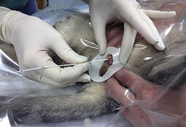
A subcutaneous ureteral bypass (SUB) device bypasses the diseased ureter entirely. A nephrostomy catheter is placed in the renal pelvis and connected to a cystostomy catheter through a subcutaneous access port, creating an alternative route for urine drainage directly from the kidney to the bladder¹˒⁴.
In many referral centres, the SUB device has become the preferred option for benign feline ureteral obstruction, particularly when ureteroliths, severe strictures or multiple ureteral lesions make stent placement difficult¹˒⁴.
Which Patients Are Good Candidates?
Referral should be considered when a cat has:
- Complete or progressive ureteral obstruction.
- Hydronephrosis with declining renal function.
- Bilateral ureteral obstruction or obstruction affecting a solitary functioning kidney.
- Ureteroliths unlikely to pass spontaneously.
- Failed medical management or persistent obstruction despite supportive care¹˒⁴.
Early referral is essential because prolonged obstruction significantly reduces the likelihood of renal recovery.
Clinical Pearl: Waiting for persistent azotemia to worsen before referral may reduce the chance of preserving renal function.
What Does Evidence Say?
Large retrospective studies have demonstrated encouraging outcomes following SUB placement. In one multicentre study involving 81 cats with benign ureteral obstruction, SUB placement achieved successful urinary diversion in most patients, with favourable long-term outcomes despite recognised postoperative complications⁴.
Another retrospective study evaluating 24 cats found that although perioperative and long-term complications—including device occlusion, urinary tract infection and the need for revision surgery—can occur, overall survival following successful SUB placement was good³.
Recent evidence also supports the use of SUB devices and ureteral stenting in selected cats with malignant urinary outflow obstruction, providing meaningful urinary decompression and improving quality of life when curative treatment is not feasible¹.
Remember: SUB Devices Require Lifelong Monitoring
Placement of a SUB device is not the end of treatment. Long-term success depends on regular postoperative surveillance.
Follow-up typically includes:
- Periodic flushing of the SUB access port.
- Ultrasonographic assessment of renal pelvis size.
- Monitoring serum creatinine and renal function.
- Urinalysis and urine culture to detect infection.
- Imaging to evaluate device patency and identify mineralisation or obstruction¹˒³˒⁴.
Owner education is equally important, as these patients require lifelong monitoring and periodic maintenance.
Take-Home Message
SUB devices and ureteral stents have fundamentally changed the management of feline ureteral obstruction. Rather than attempting technically demanding ureteral surgery, veterinarians now have minimally invasive options that offer excellent opportunities to preserve renal function and improve long-term survival. The greatest determinant of success, however, remains early recognition and timely referral. Cats with suspected ureteral obstruction should be identified promptly, stabilised ap propriately and referred before irreversible renal damage occurs.
References (Vancouver)
- Covo MS, Berent AC, Weisse CW. Use of the subcutaneous ureteral bypass device and urethral stenting for treatment of malignant urinary outflow tract obstructions in cats. J Feline Med Surg. 2024;26(9):1098612X241262666. doi:10.1177/1098612X241262666. https://pmc.ncbi.nlm.nih.gov/articles/PMC11459477/
- Butty EM, Labato MA. Subcutaneous ureteral bypass device placement with intraoperative ultrasound guidance, with or without microsurgical ureterotomy, in 24 cats. J Feline Med Surg Open Rep. 2021;7(1). https://journals.sagepub.com/doi/10.1177/1098612X211002014
- Vrijsen E, Devriendt N, Mortier F, et al. Complications and survival after subcutaneous ureteral bypass device placement in 24 cats: a retrospective study (2016–2019). J Feline Med Surg. 2021;23(9):853-862. https://doi.org/10.1177/1098612X20975374
- Wuillemin F, Vachon C, Beauchamp G, Dunn M. Subcutaneous ureteral bypass device placement in 81 cats with benign ureteral obstruction (2013–2018). J Vet Intern Med. 2021;35(6):2778-2786. doi:10.1111/jvim.16280. Available from: https://onlinelibrary.wiley.com/doi/full/10.1111/jvim.16280
Related Contents

Upcoming Event
African Swine Fever: Diagnosis and Management
African swine fever can cause severe disease and significant losses in affected pig populations.&nbs...
.jpg)
Video
Transjugular Removal of Canine Heartworms: A Clinical and Surgical Overview
Transjugular removal of canine heartworms is a specialized intervention used in selected dogs with s...

Article
Feline Pregnancy Endocrinology: Hormonal Changes and Placental Function
Pregnancy in cats involves a distinctive endocrine pattern in which ovarian and placental hormones w...

Article
Hormonal Control of Pregnancy Termination in Cats
Pregnancy maintenance in cats depends strongly on coordinated hormonal support, particularly progest...

Article
Feline Embryo Implantation and Early Placental Development
Pregnancy establishment in cats involves a sequence of precisely timed events between fertilization,...

Article
Ultrasonographic Monitoring of Pregnancy in Cats: A Gestational Timeline
Ultrasonography provides a practical way to follow the progression of feline pregnancy by identifyin...

Article
Feline Mismating: Pregnancy Diagnosis and Clinical Management
Mismating or unplanned mating is a common clinical problem in sexually mature queen cats because of...

Article
Medical Termination of Pregnancy in Queens: Drugs, Timing and Treatment Protocols
Medical termination of pregnancy (MTP) in queen cats should be approached according to the timing of...