Article
Chronic Canine Otitis: Recognising the Red Flags Before It Becomes End-Stage Ear Disease
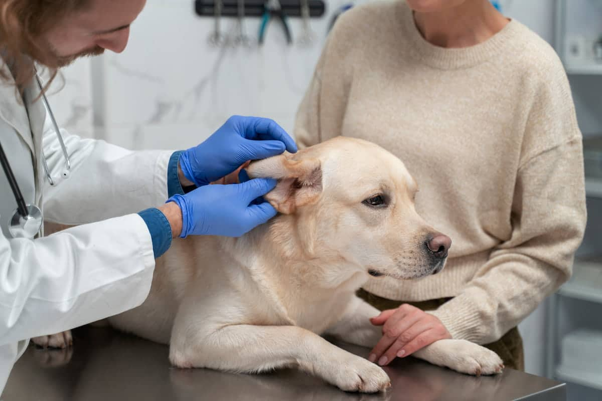
Most chronic ear cases don't become severe overnight.
They progress gradually—often because inflammation is underestimated, follow-up is delayed, or underlying disease remains untreated.
By the time fibrosis and stenosis develop, medical management becomes significantly more challenging.
Chronic Inflammation Changes the Ear Canal
Persistent inflammation causes structural changes that make future infections more likely.
These include1,2:
- Glandular hyperplasia
- Excessive cerumen production
- Hyperkeratosis
- Fibrosis
- Canal stenosis
- Mineralization in advanced disease
These changes create an ideal environment for bacteria and yeast, resulting in a cycle of chronic infection and inflammation.
Hearing Loss May Be Reversible
Owners occasionally report that their dog has become less responsive to commands during an ear infection.
Conductive hearing loss may occur because of1,3:
- Cerumen accumulation
- Purulent discharge
- Swollen ear canals
- Canal narrowing
In many patients, hearing improves considerably once the ear canal is cleaned and inflammation resolves.
Not every case of hearing loss represents permanent neurological damage.
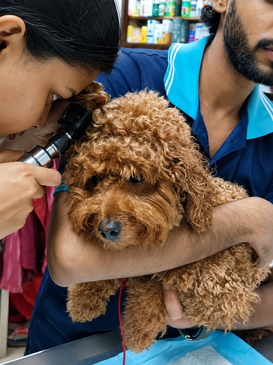
When Should You Suspect Otitis Media?
Middle ear involvement is more common than many clinicians realise, particularly in chronic cases.
Consider otitis media when you observe1:
- Recurrent otitis despite appropriate treatment
- Persistent purulent discharge
- Neurological abnormalities
- Head tilt
- Facial nerve deficits
- Nystagmus
- Poor response to therapy
Remember:
An intact tympanic membrane does not rule out middle ear disease.
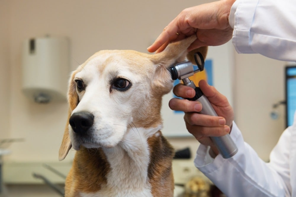
Imaging Has Its Place
Routine imaging isn't necessary for uncomplicated otitis externa.
However, advanced cases may benefit from imaging when1:
- Otitis media is suspected
- Ear canal masses are present
- Surgery is being considered
- Chronic disease fails to respond appropriately
CT imaging provides superior assessment of the tympanic bulla compared with conventional radiography.
Don't Wait Too Long to Refer
Referral should be considered when dogs present with:
- Chronic otitis
- Recurrent infections
- Severe canal stenosis
- Suspected otitis media
- Persistent Pseudomonas infections
- Cases requiring video otoscopy or deep ear flushing
Early referral often prevents irreversible ear canal damage and improves long-term patient outcomes.
Client Education Is Preventive Medicine
Many owners stop treatment as soon as scratching decreases.
Explain that:
- Clinical improvement does not equal microbiological cure.
- Follow-up cytology is essential.
- Recurrence usually indicates an unresolved underlying cause rather than treatment failure.
Setting expectations early greatly improves compliance and reduces recurrence.
Clinical Takeaway
The longer inflammation persists, the harder otitis becomes to treat.
Early diagnosis, routine cytology, appropriate ear cleaning, investigation of underlying disease, and timely referral can prevent many dogs from progressing to chronic, irreversible ear disease.
Reference
- Bajwa J. Canine otitis externa—Treatment and complications. The Canadian Veterinary Journal. 2019 Jan;60(1):97. https://pmc.ncbi.nlm.nih.gov/articles/PMC6294027/pdf/cvj_01_97.pdf
- Nuttall T. Managing recurrent otitis externa in dogs: what have we learned and what can we do better?. Journal of the American Veterinary Medical Association. 2023 Jun 1;261(S1):S10-22. https://www.research.ed.ac.uk/files/332390919/JAVMA_otitis_review_2022_unformatted_version_2.pdf
- Secker B, Shaw S, Atterbury RJ. Pseudomonas spp. in canine otitis externa. Microorganisms. 2023 Oct 28;11(11):2650. https://www.mdpi.com/2076-2607/11/11/2650
Related Contents
.jpg)
Upcoming Event
Bovine Tuberculosis: Diagnostic Challenges and Pathological Features
Bovine tuberculosis is a chronic infectious disease that continues to impact cattle health, farm eco...
Upcoming Event
Decoding the Heart in Small Animals: Session 2
Understanding cardiovascular physiology is essential for interpreting normal and abnormal cardiac fu...
.jpg)
Upcoming Event
Holistic Healing of Pets Through Homeopathy
Pet owners are increasingly interested in holistic approaches that support the overall wellbeing of...
Article
Chronic Canine Otitis: Is Culture Report Telling the Whole Story?
When a chronic otitis case doesn't respond despite selecting antibiotics based on culture and se...

Article
Chronic Canine Otitis: Is Culture Report Telling the Whole Story?
When a chronic otitis case doesn't respond despite selecting antibiotics based on culture and se...

Upcoming Event
Application of Advanced Techniques in Precision Animal Feeding
Precision feeding is transforming modern livestock production by improving feed efficiency, animal p...
Article
When Chronic Otitis Doesn't Respond: Are You Missing Biofilms and Hidden Pathogens?
Every veterinarian has encountered the frustrating ear case that has received multiple ear drops, se...
Article
Is Every Ear Infection Really an Infection? Understanding Dysbiosis in Canine Otitis
For years, canine otitis has been approached with a simple question: "Which bacteria or yeast...