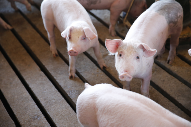
Article
Recognizing PCV2-Associated Diseases in Swine: Clinical Presentations and Pathological Findings
Porcine Circovirus type 2 (PCV2) remains an important pathogen in swine production due to its association with a broad range of clinical conditions collectively known as Porcine Circovirus-Associated Disease (PCVAD). Earlier, severe wasting conditions were mainly recognized as Post-weaning Multisystemic Wasting Syndrome (PMWS), but the understanding of PCV2-associated disease has expanded to include multiple clinical presentations affecting respiratory health, gastrointestinal function, reproduction, and skin and kidney health. The severity and expression of PCVAD depend on several interacting factors, including viral characteristics, immune status, environmental stress, and concurrent infections1.
For field veterinarians, recognizing the different manifestations of PCVAD is essential because clinical signs can vary considerably between farms. Identifying characteristic patterns and correlating them with pathological findings can help guide appropriate diagnostic and control decisions.
Post-weaning Multisystemic Wasting Syndrome (PMWS)
PMWS is one of the most recognized and severe manifestations of PCVAD, primarily affecting pigs after weaning. The condition is characterized by progressive weight loss, poor growth performance, lethargy, and increased susceptibility to secondary infections.
Common clinical observations include:
- Reduced feed intake and poor body condition
- Enlarged lymph nodes
- Respiratory difficulties
- Diarrhea
- Pallor, jaundice, and anemia in advanced cases
Affected farms may experience increased mortality, with some outbreaks showing mortality rates ranging from 30% to 40%. Gross lesions commonly include enlarged lymph nodes and pale, mottled lungs. Histopathological examination typically reveals lymphoid depletion, replacement of lymphocytes by histiocytic cells, and the presence of PCV2 within affected tissues2.
Co-infections with pathogens such as Porcine Reproductive and Respiratory Syndrome Virus (PRRSV), Mycoplasma hyopneumoniae, and swine influenza virus may worsen disease progression and contribute to increased clinical severity3.
PCV2-Associated Enteritis
PCV2-associated enteritis is an important differential diagnosis in pigs presenting with chronic diarrhea, reduced weight gain, and increased mortality, particularly between 8 and 16 weeks of age.
The intestinal lesions may resemble those caused by other enteric pathogens, making pathological confirmation important. Typical findings include granulomatous inflammation involving the small intestine, Peyer’s patches, and mesenteric lymph nodes. Extensive lymphoid depletion in Peyer’s patches and viral inclusions may also be observed5.
Because similar clinical presentations can occur with other infectious agents, veterinarians should consider PCV2-associated enteritis as part of a broader diagnostic approach rather than relying only on clinical signs.
PCV2-Associated Reproductive Failure
Reproductive problems linked with PCV2 infection have been reported in breeding herds and may present as increased numbers of mummified fetuses, non-viable piglets, and reproductive losses.
PCV2 can be detected in boar semen, and vertical transmission from sow to fetus has been demonstrated. The timing of infection during gestation influences the severity of fetal outcomes, with earlier exposure associated with more significant reproductive disruption4.
For breeding herd management, monitoring reproductive performance and considering PCV2 as a possible contributing factor may assist in identifying underlying infectious causes of reproductive failure.
Porcine Dermatitis and Nephropathy Syndrome (PDNS)
PDNS is characterized by distinctive skin lesions and kidney involvement. Affected pigs commonly develop raised purple skin lesions that may progress into red or purple scabs. Systemic vasculitis, glomerulonephritis, and dermal necrosis are characteristic pathological findings.
Clinically, affected animals may show fever, lethargy, and reduced appetite. Kidney lesions often include tan, waxy kidneys with petechial hemorrhages and multifocal glomerulonephritis, particularly within the renal cortex4.
Practical Clinical Insights
When evaluating suspected PCVAD cases, veterinarians should consider:
- The age group affected and the predominant clinical pattern within the herd.
- The possibility of concurrent infections influencing disease severity.
- The need to correlate clinical findings with characteristic tissue lesions and PCV2 detection.
- Herd-level factors such as immunity, management practices, and environmental stressors.
Recognizing the diverse clinical expressions of PCV2 infection allows veterinarians to move beyond identifying individual signs and develop a more complete understanding of disease patterns at the farm level. Early recognition of PCVAD manifestations can support timely diagnostic evaluation and implementation of appropriate control strategies.
References
- Gillespie J, Opriessnig T, Meng XJ, Pelzer K, Buechner-Maxwell V. Porcine circovirus type 2 and porcine circovirus-associated disease. Journal of veterinary internal medicine. 2009 Nov;23(6):1151-63. https://onlinelibrary.wiley.com/doi/pdf/10.1111/j.1939-1676.2009.0389.x
- Maity HK, Samanta K, Deb R, Gupta VK. Revisiting porcine circovirus infection: recent insights and its significance in the piggery sector. Vaccines. 2023 Jul 31;11(8):1308. https://www.mdpi.com/2076-393X/11/8/1308
- Grau-Roma L, Crisci E, Sibila M, Lopez-Soria S, Nofrarias M, Cortey M, Fraile L, Olvera A, Segalés J. A proposal on porcine circovirus type 2 (PCV2) genotype definition and their relation with postweaning multisystemic wasting syndrome (PMWS) occurrence. Veterinary microbiology. 2008 Apr 1;128(1-2):23-35. https://hal.science/hal-00532321v1/file/PEER_stage2_10.1016%252Fj.vetmic.2007.09.007.pdf
- Borah D, Hazarika R, Hazarika G, Saikia DP, Mili P, Bappu HP, Islam I, Barua J, Brahma D, George S. Porcine circovirus diseases: current insights and future strategies for effective Control, with a focus on Porcine circovirus 2 (PCV2). Microbiol Res J Int. 2024 Dec 30;34(12):283-98. https://www.researchgate.net/publication/387723361
Related Contents
Article
Camel Tuberculosis: Clinical Signs and Pathological Lesions
A camel may appear clinically normal despite having substantial pulmonary lesions. Conve...
.jpg)
Upcoming Event
World Animal Day
World Animal Day highlights the importance of protecting animal health and welfare through responsib...
Video
Rabies: Epitome of the One Health Paradigm
Rabies exemplifies the One Health approach, linking animal health, human health, veterinary services...
Video
One Health Webinar on Important Updates on Rabies for Veterinary Practitioners and Students
Rabies remains a major zoonotic disease requiring vigilance across veterinary practice and public he...
.jpg)
Video
Transjugular Removal of Canine Heartworms: A Clinical and Surgical Overview
Transjugular removal of canine heartworms is a specialized intervention used in selected dogs with s...

Article
Feline Pregnancy Endocrinology: Hormonal Changes and Placental Function
Pregnancy in cats involves a distinctive endocrine pattern in which ovarian and placental hormones w...

Article
Hormonal Control of Pregnancy Termination in Cats
Pregnancy maintenance in cats depends strongly on coordinated hormonal support, particularly progest...

Article
Feline Embryo Implantation and Early Placental Development
Pregnancy establishment in cats involves a sequence of precisely timed events between fertilization,...