Article
Colonoscopy-Guided Diagnosis and Medical Management of Severe Colitis in a German Shepherd Dog
Canine colitis remains one of the more challenging manifestations of inflammatory bowel disease (IBD) in small animal practice, particularly when patients present with persistent hematochezia and dyschezia despite symptomatic therapy. While many cases are initially approached as dietary or infectious enteropathies, chronic large bowel inflammation often requires a more structured diagnostic workup to reach a definitive diagnosis and guide long-term management1. The following case highlights how colonoscopy, combined with histopathology and targeted medical therapy, helped achieve complete clinical recovery in a severely affected dog.
A Challenging Large Bowel Case
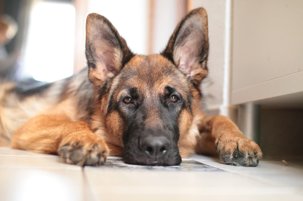
A three-year-old intact German Shepherd bitch weighing approximately 20 kg was presented with a two-week history of inappetence, straining during defecation, and passage of fresh blood in the feces. Clinical examination revealed a dull and mildly dehydrated patient with abdominal pain on palpation. Digital rectal examination demonstrated thickening of the rectal mucosa along with scant feces mixed with blood and mucus, pointing strongly toward large bowel involvement1.
Colitis in dogs may occur in several forms, including plasmacytic-lymphocytic colitis, histiocytic colitis commonly reported in Boxers, parasitic colitis, infectious colitis, ischemic colitis, and immune-mediated disease. Regardless of etiology, common presenting signs include1:
- Hematochezia
- Tenesmus
- Mucoid feces
- Dyschezia
- Constipation
In this patient, hematological examination revealed leukocytosis with neutrophilia, supporting an active inflammatory process, while serum biochemistry remained within normal limits. Fecal examination ruled out parasitic involvement, an important step before considering immunosuppressive therapy.
Imaging and Colonoscopic Findings
Abdominal radiography demonstrated gas-filled intestinal loops, while ultrasonography revealed marked thickening of the colonic wall. The mucosal wall thickness measured approximately 1.10 cm, suggesting severe inflammatory involvement of the colon. These ultrasonographic findings strongly supported large bowel pathology and justified further endoscopic evaluation1.
Although ultrasonography serves as a valuable non-invasive screening tool, definitive diagnosis of inflammatory bowel disease requires visualization and tissue sampling. As emphasized by Rychlik et al. (2007) and Rychlik and Kaczmar (2020), colonoscopy with endoscopically guided biopsy remains the cornerstone for confirming chronic inflammatory disease of the colon2,3.
The patient was prepared with 18–24 hours of fasting followed by phosphate enema administration to ensure complete evacuation of fecal material, a critical step for optimal visualization. Under sedation with xylazine and ketamine, colonoscopy was performed using a flexible video fiberscope1.
The procedure revealed severe hyperaemic ulceration and marked mucosal thickening, particularly at the transverse colon. Endoscopic-guided biopsy samples were collected for histopathological examination, which demonstrated moderate mononuclear cell infiltration consistent with inflammatory colitis1.
Practical Treatment Approach and Clinical Outcome1
The dog was initially stabilized with injectable enrofloxacin, prednisolone, and pantoprazole. Oral therapy was subsequently continued for one week using:
- Enrofloxacin
- Prednisolone
- Metronidazole
- Mesalazine
- Pantoprazole
The inclusion of mesalazine alongside corticosteroid therapy appeared particularly beneficial in controlling mucosal inflammation and accelerating recovery.
Within one week, leukocyte counts normalized and clinical signs resolved completely. Most notably, follow-up over eight months revealed no recurrence of colitis, highlighting the effectiveness of early definitive diagnosis combined with aggressive medical management.
Clinical Takeaways for Practitioners
Canine inflammatory bowel disease often presents as a diagnostic challenge because clinical signs overlap with infectious, parasitic, dietary, and neoplastic disorders (Malewska et al., 2011). This case reinforces several practical points for clinicians:
- Persistent hematochezia and tenesmus warrant detailed large bowel evaluation.
- Ultrasonography can help identify abnormal colonic thickening but cannot replace biopsy.
- Proper bowel preparation is essential for successful colonoscopy.
- Colonoscopic biopsy remains the gold standard for confirming inflammatory bowel disease.
- Combination therapy using corticosteroids and mesalazine may provide excellent clinical outcomes in severe colitis cases.
For veterinarians managing chronic enteropathies, this case demonstrates how integrating advanced diagnostics with targeted anti-inflammatory therapy can significantly improve prognosis and reduce recurrence in canine colitis.
References
- Kalaiyarasan V, Kumar SG, Sumathi D, Ruthrakumar R, Ravi R. Colonoscopic diagnosis and management of colitis in a dog. Pharma Innovation J. 2023;12(7):1093-5. https://krishikosh.egranth.ac.in/server/api/core/bitstreams/af5b1714-dee3-4b76-a8c2-eb47b544bd02/content
- Rychlik A, Nieradka R, Kander M, Depta A, Nowicki M, Sarti K. Usefulness of endoscopic examination for the diagnosis of inflammatory bowel disease in the dog. A case report. Pol J Vet Sci. 2007;10(2):113-8. https://www.researchgate.net/profile/Marcin-Nowicki-2/publication/5960110_Usefulness_of_endoscopie_examination_for_the_diagnosis_of_inflammatory_bowel_disease_in_the_dog/links/0fcfd512faaa72065a000000/Usefulness-of-endoscopie-examination-for-the-diagnosis-of-inflammatory-bowel-disease-in-the-dog.pdf
- Rychlik A, Kaczmar E. Endoscopic biopsies and histopathological findings in diagnosing chronic gastrointestinal disorders in dogs and cats. Veterinary medicine international. 2020;2020(1):8827538. https://onlinelibrary.wiley.com/doi/pdf/10.1155/2020/8827538
Related Contents
.jpg)
Upcoming Event
World Animal Day
World Animal Day highlights the importance of protecting animal health and welfare through responsib...
.jpg)
Video
Transjugular Removal of Canine Heartworms: A Clinical and Surgical Overview
Transjugular removal of canine heartworms is a specialized intervention used in selected dogs with s...

Article
Feline Pregnancy Endocrinology: Hormonal Changes and Placental Function
Pregnancy in cats involves a distinctive endocrine pattern in which ovarian and placental hormones w...

Article
Hormonal Control of Pregnancy Termination in Cats
Pregnancy maintenance in cats depends strongly on coordinated hormonal support, particularly progest...

Article
Feline Embryo Implantation and Early Placental Development
Pregnancy establishment in cats involves a sequence of precisely timed events between fertilization,...

Article
Ultrasonographic Monitoring of Pregnancy in Cats: A Gestational Timeline
Ultrasonography provides a practical way to follow the progression of feline pregnancy by identifyin...

Article
Feline Mismating: Pregnancy Diagnosis and Clinical Management
Mismating or unplanned mating is a common clinical problem in sexually mature queen cats because of...

Article
Medical Termination of Pregnancy in Queens: Drugs, Timing and Treatment Protocols
Medical termination of pregnancy (MTP) in queen cats should be approached according to the timing of...