Article
Helicobacter Suis and Gastric Microbiota in Porcine Gastric Ulceration
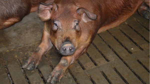
The development of gastric ulcers in pigs is influenced by multiple interacting factors, and the gastric microbiota has emerged as an important component of this process. Among the microorganisms associated with porcine gastric health, Helicobacter suis has received particular attention because of its ability to colonize the gastric mucosa and alter the stomach environment. Although gastric ulceration cannot be attributed to a single infectious agent, changes in gastric acid secretion and microbial populations may contribute to lesion development and progression1. Understanding these microbial interactions enables veterinarians to interpret gastric lesions more comprehensively while considering other contributing nutritional and management factors.
Helicobacter suis Colonization in the Pig Stomach
Helicobacter suis is a zoonotic bacterium that colonizes the gastric mucosa of pigs worldwide1. Infection becomes increasingly common with age. Colonization has been reported at low levels in suckling pigs, increasing progressively in weaned pigs, slaughter-age pigs, and adult animals. High prevalence in adult pigs suggests that the host immune response does not readily eliminate the organism1,2.
Colonization patterns also appear to change over time. In younger pigs, H. suis is predominantly found within the pyloric gland region, whereas prolonged infection is associated with greater bacterial numbers in the fundal gland region1,2. These findings indicate that bacterial distribution within the stomach may evolve as infection persists.
How Microbial Changes May Influence Ulcer Development
The precise mechanism by which H. suis contributes to gastric ulceration remains incompletely understood, but alterations in gastric acid secretion appear to play an important role. During the earlier stages of infection, reduced gastric acid secretion may alter the microbial composition of the pars esophagea, creating conditions that favour the establishment of other bacterial populations2,3.
One organism receiving increasing attention is Fusobacterium gastrosuis. A higher number of this bacterium has been identified in stomachs affected by H. suis, suggesting that changes induced by H. suis may facilitate its proliferation1. Colonization of the pars esophagea by F. gastrosuis has been associated with lesion development, and metabolites produced by this organism may contribute to epithelial cell death and gastric ulcer formation4.
As infection progresses into the chronic phase, gastric acid secretion may increase, potentially worsening lesions in the pars esophagea. Because this non-glandular region lacks a protective mucus layer, it remains particularly susceptible to acid-induced injury2,3. These observations support the concept that microbial alterations and acid secretion changes may act together during ulcer development.
The Role of Gastric Microbiota
The composition of the gastric microbiota may also influence mucosal health. A reduction in beneficial Lactobacillus populations has been associated with increased erosion and ulceration4. Lower numbers of these bacteria may reduce the production of antimicrobial substances such as lactic acid and bacteriocins while increasing gastric pH, creating a more favourable environment for pathogenic organisms1.
Other microorganisms, including Arcobacter spp., Bacillus spp., Candida spp., and additional bacterial species, have occasionally been detected in pigs with gastric lesions. However, these organisms have not been consistently recognised as highly specific risk factors for gastric ulceration1.
Although the direct effect of probiotic supplementation on gastric ulcer prevention has not yet been established, the recognised role of gastric microflora suggests that maintaining a balanced microbial environment may warrant further consideration in future herd health strategies1.
Practical Clinical Insights
When gastric ulcers are identified in a herd, veterinarians should recognise that microbial factors represent only one component of a multifactorial disease process.
Practical considerations include:
- Consider H. suis as a potential contributor when evaluating gastric ulceration, particularly in older pigs.
- Interpret microbial findings alongside nutritional, housing, and management factors rather than in isolation.
- Recognise that age-related changes in bacterial colonization may influence lesion development.
- Be aware that disruption of beneficial gastric microflora, particularly Lactobacillus spp., may accompany ulcer progression.
- Remember that current evidence does not support attributing gastric ulceration to a single infectious organism.
A practical clinical approach integrates microbial findings with the overall herd picture, recognising that interactions between gastric bacteria, acid secretion, and environmental factors collectively influence the development and severity of gastric ulcers in pigs.
References
- Krepelková Z, Novotný J, Bárdová K, Link R, Csörgö A. Gastric ulcers in pigs–a review. Folia veterinaria. 2024 Jun 27;68(2):33-42. https://sciendo.com/pdf/10.2478/fv-2024-0015
- De Witte C, Devriendt B, Flahou B, Bosschem I, Ducatelle R, Smet A, Haesebrouck F. Helicobacter suis induces changes in gastric inflammation and acid secretion markers in pigs of different ages. Veterinary research. 2017 Jun 15;48(1):34. https://link.springer.com/content/pdf/10.1186/s13567-017-0441-6.pdf
- Ziegler A, Gonzalez L, Blikslager A. Large animal models: the key to translational discovery in digestive disease research. Cellular and molecular gastroenterology and hepatology. 2016 Nov 1;2(6):716-24. https://www.sciencedirect.com/science/article/pii/S2352345X16301047
- Taillieu E, Taelman S, De Bruyckere S, Goossens E, Chantziaras I, Van Steenkiste C, Yde P, Hanssens S, De Meyer D, Van Criekinge W, Stock M. The role of Helicobacter suis, Fusobacterium gastrosuis, and the pars oesophageal microbiota in gastric ulceration in slaughter pigs receiving meal or pelleted feed. Veterinary Research. 2024 Feb 5;55(1):15. https://link.springer.com/content/pdf/10.1186/s13567-024-01274-1.pdf
Related Contents

Article
The Elephant is Walking Differently: Where Should the Investigation Begin?
When an elephant begins walking differently, the first challenge is not necessarily to identify the...

Article
When an Elephant’s Blood Report Says “Abnormal”: How Should It Be Interpreted?
A laboratory report can flag a value as abnormal even when an elephant appears clinically healthy. C...
.jpg)
Upcoming Event
World Animal Day
World Animal Day highlights the importance of protecting animal health and welfare through responsib...
Video
Rabies: Epitome of the One Health Paradigm
Rabies exemplifies the One Health approach, linking animal health, human health, veterinary services...
Video
One Health Webinar on Important Updates on Rabies for Veterinary Practitioners and Students
Rabies remains a major zoonotic disease requiring vigilance across veterinary practice and public he...
.jpg)
Video
Transjugular Removal of Canine Heartworms: A Clinical and Surgical Overview
Transjugular removal of canine heartworms is a specialized intervention used in selected dogs with s...

Article
Feline Pregnancy Endocrinology: Hormonal Changes and Placental Function
Pregnancy in cats involves a distinctive endocrine pattern in which ovarian and placental hormones w...

Article
Hormonal Control of Pregnancy Termination in Cats
Pregnancy maintenance in cats depends strongly on coordinated hormonal support, particularly progest...