Article
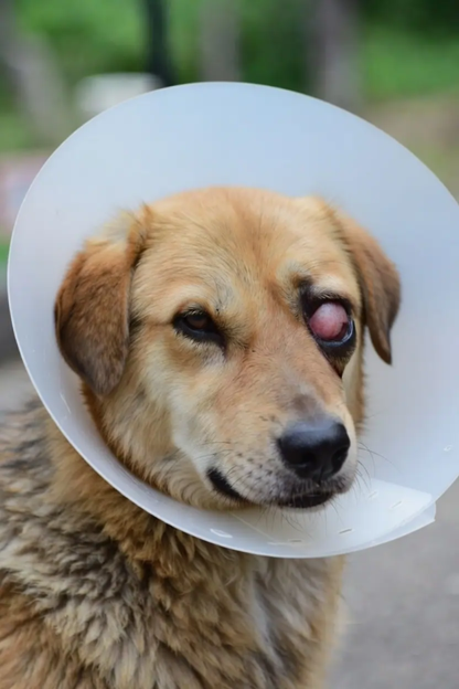
Morgan's Pocket Technique: Why It Remains the First Choice for Cherry Eye Repair
For many years, excision of the prolapsed third eyelid gland was considered a simple solution for cherry eye. However, advances in veterinary ophthalmology have fundamentally changed this approach. Today, the focus is no longer on removing the gland but on preserving its function, as the gland contributes nearly 30–50% of the aqueous component of the canine tear film. Loss of this gland significantly increases the risk of keratoconjunctivitis sicca (KCS), making gland-preserving procedures the standard of care1.
Among the various surgical techniques described over the years, Morgan's Pocket Technique has consistently emerged as the preferred first-line procedure because it combines excellent clinical outcomes with relative technical simplicity1,2.
Why Morgan's Pocket Technique remains the benchmark
Originally described by Morgan and colleagues, this technique repositions the prolapsed gland by creating two conjunctival incisions on the bulbar surface of the third eyelid and enclosing the gland within a conjunctival pocket using absorbable sutures. Unlike anchoring procedures that secure the gland to adjacent orbital tissues, the Morgan's Pocket Technique restores the gland close to its natural anatomical position while preserving its mobility and secretory function1,2.
A prospective clinical study involving 17 dogs (20 affected eyes) demonstrated an overall 94.1% surgical success rate, with recurrence observed in only one eye (5.9%). Apart from one dog that developed prolonged epiphora, postoperative complications were minimal, confirming that the procedure is both safe and reliable for routine clinical practice2.
Systematic evaluation of available surgical techniques also concludes that no alternative gland-preserving procedure has consistently demonstrated superior long-term outcomes. For uncomplicated cases, Morgan's Pocket Technique continues to represent the benchmark against which other techniques are compared1.
Choosing the right patient is equally important
Not every case of cherry eye presents under ideal conditions. Dogs with recent gland prolapse generally have healthier gland tissue, making surgical replacement easier and reducing the likelihood of postoperative complications. Chronic prolapse, on the other hand, often leads to inflammation, fibrosis, and enlargement of the gland, which can complicate repositioning and increase the risk of recurrence 1,2.
Before scheduling surgery, a complete ophthalmic examination should include:
- Assessment of the duration of prolapse
- Evaluation of conjunctival inflammation
- Fluorescein staining to identify corneal ulceration
- Schirmer Tear Test (STT) to establish baseline tear production
- Examination of the opposite eye for early or subclinical prolapse
Early surgical intervention remains one of the simplest ways to improve long-term outcomes.
Genetics explains why some breeds are at greater risk
Recent research has strengthened the understanding that cherry eye is not simply an anatomical abnormality but also has a significant genetic basis. A 2024 genome-wide association study identified the FGF4L1 retrogene insertion as a major genetic risk factor for prolapse of the nictitans gland, supporting the theory that inherited connective tissue weakness contributes to gland instability3. Earlier pedigree analyses also demonstrated that inheritance is complex rather than following a simple Mendelian pattern4.
Breeds frequently affected include:
- English Bulldog
- French Bulldog
- American Cocker Spaniel
- Beagle
- Lhasa Apso
- Shih Tzu
- Cane Corso
- Neapolitan Mastiff
Because many predisposed dogs eventually develop bilateral disease, veterinarians should educate owners that prolapse in the second eye remains a possibility even after successful surgery on the first eye3,4.
When should another technique be considered?
Although Morgan's Pocket Technique remains the preferred option for most primary cases, it may not be suitable for every patient. Dogs with recurrent prolapse, severe connective tissue laxity, or marked gland enlargement may require alternative gland-preserving procedures, such as anchoring techniques or combined pocket-and-anchoring approaches1.
Importantly, recurrence should not always be viewed as a surgical failure. Underlying breed-related connective tissue abnormalities may predispose certain dogs to recurrent prolapse despite meticulous surgical technique3,4.
Clinical Pearl
The success of cherry eye surgery depends less on the complexity of the procedure and more on preserving the biology of the gland. For most uncomplicated cases, Morgan's Pocket Technique offers an excellent combination of simplicity, gland preservation, and long-term success. Early intervention and careful patient selection remain the keys to optimal outcomes.
References
- White C, Brennan ML. An evidence-based rapid review of surgical techniques for correction of prolapsed nictitans glands in dogs. Vet Sci. 2018;5(3):75. https://pmc.ncbi.nlm.nih.gov/articles/PMC6163435/
- Deveci MZ, İşler CT, Yurtal Z, Altuğ ME, Kırgız Ö. Evaluation of Morgan's pocket technique in the treatment of nictitans gland prolapse in dogs. Turkish Journal of Veterinary & Animal Sciences. 2020;44(3):521–527. https://journals.tubitak.gov.tr/cgi/viewcontent.cgi?article=1191&context=veterinary
- Freyer J, Labadie JD, Huff JT, et al. Association of FGF4L1 Retrogene Insertion with Prolapsed Gland of the Nictitans (Cherry Eye) in Dogs. Genes. 2024;15(2):198. https://pmc.ncbi.nlm.nih.gov/articles/PMC10887708/
- O'Neill DG, Church DB, McGreevy PD, et al. Investigating the inheritance of prolapsed nictitating membrane glands in a large canine pedigree. Canine Genet Epidemiol. 2015;2:7. https://pmc.ncbi.nlm.nih.gov/articles/PMC4361898/
Related Contents

Upcoming Event
African Swine Fever: Diagnosis and Management
African swine fever can cause severe disease and significant losses in affected pig populations.&nbs...
.jpg)
Video
Transjugular Removal of Canine Heartworms: A Clinical and Surgical Overview
Transjugular removal of canine heartworms is a specialized intervention used in selected dogs with s...

Article
Feline Pregnancy Endocrinology: Hormonal Changes and Placental Function
Pregnancy in cats involves a distinctive endocrine pattern in which ovarian and placental hormones w...

Article
Hormonal Control of Pregnancy Termination in Cats
Pregnancy maintenance in cats depends strongly on coordinated hormonal support, particularly progest...

Article
Feline Embryo Implantation and Early Placental Development
Pregnancy establishment in cats involves a sequence of precisely timed events between fertilization,...

Article
Ultrasonographic Monitoring of Pregnancy in Cats: A Gestational Timeline
Ultrasonography provides a practical way to follow the progression of feline pregnancy by identifyin...

Article
Feline Mismating: Pregnancy Diagnosis and Clinical Management
Mismating or unplanned mating is a common clinical problem in sexually mature queen cats because of...

Article
Medical Termination of Pregnancy in Queens: Drugs, Timing and Treatment Protocols
Medical termination of pregnancy (MTP) in queen cats should be approached according to the timing of...