Article
Diagnosis of Suspensory Ligament Pathology in Horses
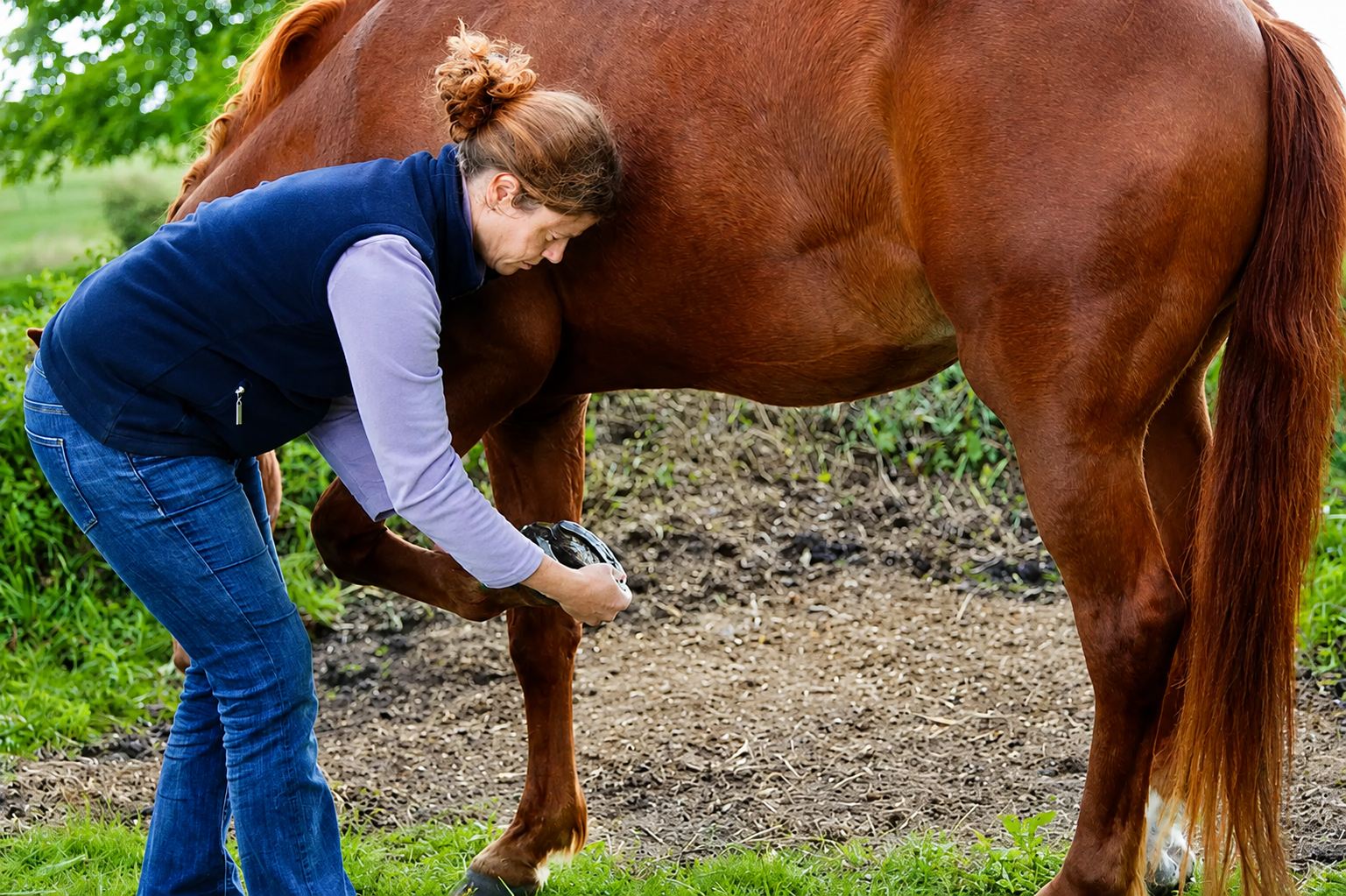
Diagnosing suspensory ligament pathology can be challenging due to the ligament's anatomical location and the lack of specific clinical signs, particularly in chronic cases. While acute injuries may present with heat, swelling, and pain on palpation, chronic lesions often lack obvious external indicators1. Consequently, diagnosis typically relies on a combination of diagnostic anaesthesia and advanced imaging techniques.
Diagnostic Anaesthesia
Localising pain originating from the suspensory ligament can be difficult because of variations in nerve supply and the proximity of surrounding joints and tendon sheaths. In the hindlimb, anaesthesia of the deep branch of the lateral plantar nerve (DBLPN) may also desensitise distal structures, including portions of the tarsal joint2.
Recent refinements in nerve-blocking techniques have shown that small-volume injections adjacent to the DBLPN can effectively anaesthetise the suspensory ligament while minimising interference with neighbouring structures. However, complete specificity remains difficult to achieve. In the forelimb, direct synovial communication between the carpometacarpal joint and the proximal suspensory ligament further complicates diagnostic analgesia3,4.
Ultrasonography: The Primary Diagnostic Tool
Ultrasonography remains the most widely used imaging modality for diagnosing SL pathology. Common ultrasonographic abnormalities include ligament enlargement, poorly defined margins, hypoechoic regions, and diffuse reductions in echogenicity, particularly within the proximal suspensory ligament1.
Lesions affecting the suspensory branches often appear as anechoic or hypoechoic areas accompanied by heterogeneous echogenicity and disruption of the normal fibrillar pattern. Interestingly, studies have demonstrated ultrasonographic abnormalities in clinically sound horses, including approximately 7% of Thoroughbred racehorses, 30% of National Hunt horses, 58% of showjumping Warmbloods, and 20% of working Quarter Horses1,5.
Advanced Ultrasonographic Techniques
The accuracy of ultrasonography can be affected by operator experience, particularly when evaluating the proximal suspensory ligament. Diagnostic accuracy improves when medial and lateral imaging approaches are combined with angle-contrast techniques and non-weightbearing examinations.
Doppler ultrasonography has shown promise by enabling assessment of blood flow changes associated with pathology and demonstrates good agreement with conventional B-mode ultrasonography6.
Elastography is another emerging technique that estimates tissue stiffness. Studies have shown that acute lesions tend to be softer than chronic lesions and that stiffness increases during healing. Although promising for monitoring disease progression, elastography has limitations in detecting small proximal lesions and has not yet become a routine clinical tool1,7.
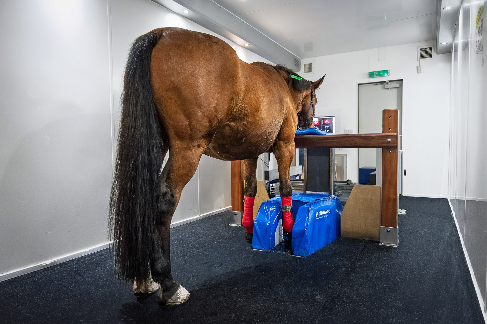
Magnetic Resonance Imaging (MRI)
MRI has significantly advanced the diagnosis of suspensory ligament injuries by providing superior soft-tissue detail and earlier detection of pathology. MRI can identify interfascicular spaces, fibre disruption, ligament thickening, adhesions, and core lesions that may not be visible on ultrasound.
Importantly, MRI can detect biochemical changes before obvious structural damage develops, potentially enabling earlier intervention and improved outcomes. However, considerable variation exists in the normal MRI appearance of the suspensory ligament, particularly proximally, requiring careful interpretation1.
MRI is especially valuable for diagnosing proximal suspensory ligament disease and branch injuries, with studies demonstrating substantially higher sensitivity than ultrasonography.
Computed Tomography and Nuclear Scintigraphy
Computed tomography (CT) can identify mineralisation, enlargement of the suspensory ligament, and associated bony abnormalities. Contrast-enhanced CT additionally allows visualisation of vascular changes within the ligament and may serve as a useful alternative when MRI is unavailable1.
Nuclear scintigraphy can assist in identifying suspensory ligament pathology, particularly when combined with radiography or ultrasonography. However, its sensitivity varies and some lesions may remain undetected1.
Conclusion
While ultrasonography remains the cornerstone of suspensory ligament diagnosis in equine practice, MRI offers superior sensitivity and earlier detection of pathology. Emerging technologies such as Doppler ultrasonography, elastography, CT, and scintigraphy continue to enhance diagnostic capabilities and may improve clinical decision-making in the future.
References
- Guest DJ, Birch HL, Thorpe CT. A review of the equine suspensory ligament: Injury prone yet understudied. Equine Veterinary Journal. 2025 Sep;57(5):1167-82. https://beva.onlinelibrary.wiley.com/doi/pdfdirect/10.1111/evj.14447
- Labens R, Schramme MC, Robertson ID, Thrall DE, Redding WR. Clinical, magnetic resonance, and sonographic imaging findings in horses with proximal plantar metatarsal pain. Veterinary radiology & ultrasound. 2010 Jan;51(1):11-8. https://doi.org/10.1111/j.1740-8261.2009.01614.x
- Contino EK, King MR, Valdés‐Martínez A, McIlwraith CW. In vivo diffusion characteristics following perineural injection of the deep branch of the lateral plantar nerve with mepivacaine or iohexol in horses. Equine veterinary journal. 2015 Mar;47(2):230-4. https://doi.org/10.1111/evj.12261
- Gerdes C, Morgan R, Terry R, Foote A, Smith R. Computed tomographic arthrography, gross anatomy and histology demonstrate a communication between synovial invaginations in the proximal aspect of the third interosseous muscle and the carpometacarpal joint in horses. Frontiers in Veterinary Science. 2022 Sep 2;9:958598. https://doi.org/10.3389/fvets.2022.958598
- Read RM, Boys-Smith S, Bathe AP. Subclinical ultrasonographic abnormalities of the suspensory ligament branches are common in elite showjumping warmblood horses. Frontiers in Veterinary Science. 2020 Mar 17;7:117. https://doi.org/10.3389/fvets.2020.00117
- Rabba S, Grulke S, Verwilghen D, Evrard L, Busoni V. B‐mode and power Doppler ultrasonography of the equine suspensory ligament branches: a descriptive study on 13 horses. Veterinary Radiology & Ultrasound. 2018 Jul;59(4):453-60. https://doi.org/10.1111/vru.12610
- Lustgarten M, Redding WR, Labens R, Davis W, Daniel TM, Griffith E, Seiler GS. Elastographic evaluation of naturally occuring tendon and ligament injuries of the equine distal limb. Veterinary Radiology & Ultrasound. 2015 Nov;56(6):670-9. https://doi.org/10.1111/vru.12284
Related Contents
.jpg)
Upcoming Event
Bovine Tuberculosis: Diagnostic Challenges and Pathological Features
Bovine tuberculosis is a chronic infectious disease that continues to impact cattle health, farm eco...

Upcoming Event
Holistic Healing of Pets Through Homeopathy
Pet owners are increasingly interested in holistic approaches that support the overall wellbeing of...
.jpg)
Upcoming Event
Neonatology: The Crucial First Two Weeks of Life
The neonatal period, particularly the first 14 days of life, is one of the most vulnerable stages in...
Article
Beyond Medication: Rehabilitation Strategies That Can Transform Outcomes in Equine Osteoarthritis
Osteoarthritis (OA) remains one of the leading causes of lameness, reduced athletic perfor...
Article
Beyond Overstrain: Understanding the Causes and Pathophysiology of Suspensory Ligament Injuries in Horses
Suspensory ligament (SL) injuries are among the most common musculoskeletal disorders affecting equi...
Article
Beyond Overstrain: Understanding the Causes and Pathophysiology of Suspensory Ligament Injuries in Horses
Suspensory ligament (SL) injuries are among the most common musculoskeletal disorders affecting equi...
Article
Current Treatment Approaches for Suspensory Ligament Injuries in Horses
Suspensory ligament (SL) injuries remain one of the most frustrating causes of lameness an...
Article
Right Dorsal Displacement in Horses: Understanding the Causes Behind a Common Surgical Colic
The equine large colon is one of the most mobile structures within the gastrointestinal tract. Becau...