Article
Pathogenesis and Clinical Progression of Gastric Ulcers in Pigs
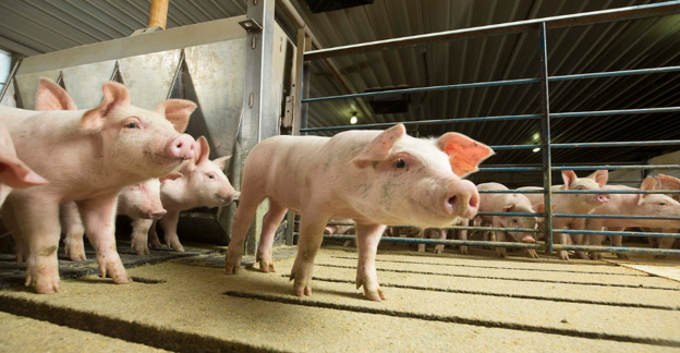
Gastric ulceration in pigs most commonly affects the pars esophagea, the non-glandular region of the stomach that lacks a protective mucus layer. This anatomical characteristic makes it particularly vulnerable when normal gastric conditions are disturbed. Ulcer formation can occur rapidly, and although many affected pigs remain clinically normal, severe lesions may lead to reduced performance, anaemia, or sudden death1. For practicing veterinarians, understanding how these lesions develop and progress is essential for recognising affected animals and identifying opportunities for timely intervention.
Why the Pars Esophagea Is Vulnerable
The porcine stomach consists of four regions: oesophageal, cardiac, fundal, and pyloric. The pars esophagea is the only glandless portion and forms a continuation of the oesophageal mucosa2. Under normal conditions, the proximal part of the stomach, including the pars esophagea and cardiac region, maintains a relatively neutral pH of 5–7 due to bicarbonate present in saliva and cardiac gland secretions. In contrast, the fundal and pyloric regions maintain a highly acidic environment with a pH of 2–3 through hydrochloric acid production by parietal cells3.
Normally, these compartments remain separated. When this segregation is disrupted, hydrochloric acid, pepsin, and bile can come into contact with the pars esophagea. Because this region lacks a protective mucus barrier, repeated exposure leads to irritation, inflammation, keratosis, erosion, and eventually ulcer formation3.
Progression from Healthy Tissue to Ulceration
Gastric ulceration develops through a predictable sequence of pathological changes. Initially, the pars esophagea appears smooth, white, and healthy. Continuous irritation stimulates thickening of the epithelial surface, producing parakeratosis or keratosis. As the damaging effects of gastric contents persist, superficial erosions develop and progressively extend into deeper tissue layers. Without resolution of the underlying cause, these erosions progress to ulcers. If conditions improve, healing occurs through scar formation, although fibrosis may narrow the oesophageal opening and result in stenosis4.
The progression can be summarised as:
- Healthy pars esophagea
- Keratosis
- Erosion
- Ulcer
- Scar formation4
Ulcers vary considerably in size, depth, and number. Extensive lesions may involve the entire pars esophagea, while healed ulcers often leave puckered or star-shaped scars1.
Clinical Progression and Associated Signs
Disease progression varies according to lesion severity and duration. Ulcers may develop within less than 24 hours and, in some cases, within only 12 hours. Many pigs remain subclinically affected despite having extensive erosive lesions1.
Peracute and acute cases may show no warning signs before sudden death caused by severe gastric haemorrhage or perforation leading to fatal peritonitis1. In subacute or chronic cases, clinical signs become more apparent and may include:
- Reduced feed intake and growth performance
- Pallor of the mucous membranes
- Weakness and reluctance to walk
- Recumbency
- Anorexia
- Melena
- Anaemia with reduced haematocrit, haemoglobin, and erythrocyte count
- Teeth grinding associated with gastric pain
- Greenish bile-stained vomiting1
At necropsy, lesions are typically confined to the pars esophagea. Early lesions appear thickened, raised, and yellowish due to exposure to bile and hydrochloric acid. Continued erosion creates crater-like ulcers that may damage major blood vessels, resulting in significant haemorrhage1.
Practical Clinical Insights
Recognition of ulcer progression is valuable when evaluating pigs with unexplained reductions in performance or unexpected mortality.
Important clinical considerations include:
- Remember that severe gastric lesions may be present even in pigs showing minimal or no clinical signs.
- Consider gastric ulceration in cases of unexplained anaemia, melena, poor growth, or sudden death.
- Recognise that lesion progression can occur rapidly, making early identification of herd-level risk factors particularly important.
- During post-mortem examination, carefully inspect the pars esophagea, as lesions are typically localised to this region and may vary considerably in severity.
A thorough understanding of the sequence from mucosal irritation to ulcer formation enables veterinarians to better interpret clinical findings and pathological lesions while supporting timely herd health interventions.
References
- Krepelková Z, Novotný J, Bárdová K, Link R, Csörgö A. Gastric ulcers in pigs–a review. Folia veterinaria. 2024 Jun 27;68(2):33-42. https://sciendo.com/pdf/10.2478/fv-2024-0015
- Gandarillas M, Bas F. The domestic pig (Sus scrofa domestica) as a model for evaluating nutritional and metabolic consequences of bariatric surgery practiced on morbid obese humans. Ciencia e investigación agraria. 2009 Aug;36(2):163-76. https://www.scielo.cl/pdf/ciagr/v36n2/art02.pdf
- Haesebrouck F, Pasmans F, Flahou B, Chiers K, Baele M, Meyns T, Decostere A, Ducatelle R. Gastric helicobacters in domestic animals and nonhuman primates and their significance for human health. Clinical microbiology reviews. 2009 Apr;22(2):202-23. https://pmc.ncbi.nlm.nih.gov/articles/PMC2668234/pdf/0041-08.pdf
- Peralvo-Vidal JM, Weber NR, Nielsen JP, Bache JK, Haugegaard S, Pedersen AØ. Risk factors for gastric ulceration in nursery pigs. Preventive veterinary medicine. 2021 Apr 1;189:105298. https://doi.org/10.1016/j.prevetmed.2021.105298
Related Contents

Article
The Elephant is Walking Differently: Where Should the Investigation Begin?
When an elephant begins walking differently, the first challenge is not necessarily to identify the...

Article
When an Elephant’s Blood Report Says “Abnormal”: How Should It Be Interpreted?
A laboratory report can flag a value as abnormal even when an elephant appears clinically healthy. C...
.jpg)
Upcoming Event
World Animal Day
World Animal Day highlights the importance of protecting animal health and welfare through responsib...
Video
Rabies: Epitome of the One Health Paradigm
Rabies exemplifies the One Health approach, linking animal health, human health, veterinary services...
Video
One Health Webinar on Important Updates on Rabies for Veterinary Practitioners and Students
Rabies remains a major zoonotic disease requiring vigilance across veterinary practice and public he...
.jpg)
Video
Transjugular Removal of Canine Heartworms: A Clinical and Surgical Overview
Transjugular removal of canine heartworms is a specialized intervention used in selected dogs with s...

Article
Feline Pregnancy Endocrinology: Hormonal Changes and Placental Function
Pregnancy in cats involves a distinctive endocrine pattern in which ovarian and placental hormones w...

Article
Hormonal Control of Pregnancy Termination in Cats
Pregnancy maintenance in cats depends strongly on coordinated hormonal support, particularly progest...