Article
Recognizing Feline Atopic Skin Syndrome: Key Clinical Reaction Patterns
When a cat presents with pruritus, alopecia, crusting or ulcerative lesions, the visible skin changes may point toward an allergic process, but they do not automatically establish feline atopic skin syndrome (FASS). Cats show allergic skin disease through a range of cutaneous reaction patterns, and several nonallergic conditions can produce the same appearances. For the practicing veterinarian, recognizing these patterns is therefore the first step toward building an appropriate diagnostic pathway1.
Four Patterns That Should Raise Suspicion
The major cutaneous reaction patterns associated with FASS are miliary dermatitis (MD), self-induced alopecia/hypotrichosis (SIAH), head and neck pruritus (HNP), and eosinophilic granuloma complex (EGC). They may occur individually or in combination. Their presence, however, becomes meaningful for FASS only after other possible causes have been excluded1.
Miliary Dermatitis: Examine Beyond the Coat
Miliary dermatitis typically appears as numerous small papules, approximately 1–2 mm, usually surrounded by crusts. The lesions may be localized or generalized. Pruritus is usually present, and excoriations, erosions and varying degrees of hair loss may develop as a consequence1.
Do not dismiss MD simply because the owner reports that the cat is not scratching. Some cats may genuinely be nonpruritic, while in others the grooming or scratching behaviour may simply go unnoticed. In these cases, the cat can appear clinically normal from a distance, with the lesions becoming apparent only when the skin is examined closely2.
Self-Induced Alopecia: Look for the Cause of Hair Loss3
SIAH is another important pattern. Cats may remove their own hair through excessive grooming, making the condition appear to be spontaneous alopecia. This is particularly relevant when a behavioural explanation is considered early in the assessment.
In one group of 21 cats referred for evaluation of presumed psychogenic alopecia, a primary behavioural or psychogenic cause was demonstrated in only two cats. Sixteen had pruritic dermatitis alone, while three had pruritic disease with a superimposed behavioural component.
The practical message is important: hair loss in a cat should not be labelled psychogenic before pruritic skin disease has been adequately investigated.
Head and Neck Pruritus: Assess the Extent of Self-Trauma
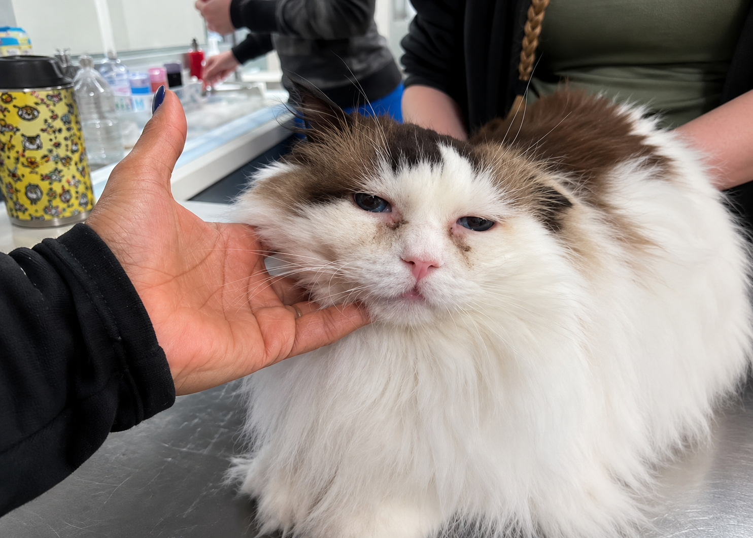
HNP is characterized by often intense pruritus affecting the face, head, and neck. Cats may claw or scratch these areas vigorously, producing excoriation, erosion, and ulceration. Blepharitis can occur, with or without corneal ulceration. When self-trauma becomes severe, protective measures such as bandages or collars may be required to reduce further damage1.
Eosinophilic Granuloma Complex: Recognize Its Different Presentations
EGC includes several clinical syndromes. Indolent ulcer, also known as “rodent ulcer,” typically affects the upper lip at or near the mucocutaneous junction. It begins as focal ulceration and may progress to extensive ulceration and fibrosis. It is generally not pruritic unless complicated by bacterial infection3.
Eosinophilic granuloma, or linear granuloma, may present as linear dermal thickening on the caudal hindlimbs. Lesions can also occur in the mouth, particularly on the tongue or hard palate, or appear as poorly defined chin swelling. These lesions may or may not be pruritic3.
Eosinophilic plaques are most frequently found on the ventral abdomen and medial thighs. They are raised and often eroded or ulcerated, with lesions ranging from circular to oval or serpiginous. Intense pruritus may drive further self-trauma and perpetuate inflammation, and secondary bacterial infection can complicate these lesions3.
How Often Do These Patterns Occur?
Among 263 cats diagnosed exclusively with FASS, the reported prevalence was 31.2% for MD, 60.1% for SIAH, 43.0% for HNP and 25.9% for one or more forms of EGC. Multiple patterns were common, with 37.7% of cats reported to have at least two syndromes1,3.
The distribution of lesions can also provide useful clinical context. Frequently affected sites include the face, head, neck, and pinnae, ventral abdomen, medial aspects of the legs, and dorsum. Paw involvement appears to be uncommon compared with these locations1,4.
Pattern Recognition Is Only the Beginning
The same reaction patterns may occur with flea allergy, food allergy, parasitic disease, dermatophytosis, bacterial or Malassezia-associated disease and other dermatological disorders. Therefore, a compatible pattern should prompt further investigation rather than an immediate diagnosis of FASS1.
For everyday practice, the most useful approach is simple: identify the reaction pattern, document its distribution and severity, assess self-trauma and then work through the relevant differentials. Recognizing what the skin is expressing can make the next diagnostic step clearer—but the pattern itself is not the diagnosis.
References
- Hobi S, Linek M, Marignac G, Olivry T, Beco L, Nett C, Fontaine J, Roosje P, Bergvall K, Belova S, Koebrich S. Clinical characteristics and causes of pruritus in cats: a multicentre study on feline hypersensitivity‐associated dermatoses. Veterinary dermatology. 2011 Oct;22(5):406-13. https://www.zora.uzh.ch/server/api/core/bitstreams/e17e1a30-a25f-4b99-9ad0-78e080724b1a/content
- Buckley L. Treatment of presumed allergic skin disease in cats. In Practice. 2017 Jun;39(6):242-54. https://livrepository.liverpool.ac.uk/3008504/1/Challenge%20of%20treating%20FASD%20proof.pdf
- Santoro D, Pucheu‐Haston CM, Prost C, Mueller RS, Jackson H. Clinical signs and diagnosis of feline atopic syndrome: detailed guidelines for a correct diagnosis. Veterinary dermatology. 2021 Feb;32(1):26-e6. https://onlinelibrary.wiley.com/doi/pdfdirect/10.1111/vde.12935
- Scott DW, Miller WH. Feline atopic dermatitis: a retrospective study of 194 cases (1988–2003). Jpn J Vet Dermatol. 2013 Nov 13;19(3):135-47. https://scholar.archive.org/work/rfaelwvjffcpfol7pdgord66lu/access/wayback/https://www.jstage.jst.go.jp/article/jjvd/19/3/19_135/_pdf
Related Contents
.jpg)
Video
Transjugular Removal of Canine Heartworms: A Clinical and Surgical Overview
Transjugular removal of canine heartworms is a specialized intervention used in selected dogs with s...

Article
Feline Pregnancy Endocrinology: Hormonal Changes and Placental Function
Pregnancy in cats involves a distinctive endocrine pattern in which ovarian and placental hormones w...

Article
Hormonal Control of Pregnancy Termination in Cats
Pregnancy maintenance in cats depends strongly on coordinated hormonal support, particularly progest...

Article
Feline Embryo Implantation and Early Placental Development
Pregnancy establishment in cats involves a sequence of precisely timed events between fertilization,...

Article
Ultrasonographic Monitoring of Pregnancy in Cats: A Gestational Timeline
Ultrasonography provides a practical way to follow the progression of feline pregnancy by identifyin...

Article
Feline Mismating: Pregnancy Diagnosis and Clinical Management
Mismating or unplanned mating is a common clinical problem in sexually mature queen cats because of...

Article
Medical Termination of Pregnancy in Queens: Drugs, Timing and Treatment Protocols
Medical termination of pregnancy (MTP) in queen cats should be approached according to the timing of...

Article
Safety and Aftercare Following Pregnancy Termination in Queen Cats
Pregnancy termination in queen cats requires more than selecting an appropriate abortifacient. Once...