Article
Beyond Navicular Disease: Decoding Distal Border Lesions in Horses
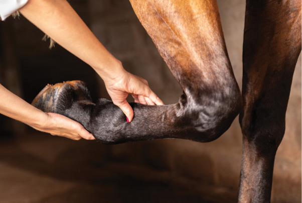
Forelimb lameness can be a diagnostic challenge, and the navicular bone is often at the center of the investigation. Although small, this boat-shaped bone plays a critical role in force distribution within the equine foot and serves as a fulcrum for the deep digital flexor tendon (DDFT) during movement1.
When excessive biomechanical stress overwhelms the foot's ability to adapt, pathological changes can develop within the navicular apparatus, leading to chronic palmar foot pain and reduced athletic performance.
Look Beyond the Bone
Navicular syndrome is no longer considered a disease of the navicular bone alone. Advanced imaging has shown that lesions frequently involve the DDFT, distal sesamoidean impar ligament (DSIL), navicular bursa, and surrounding soft tissues.
In horses with chronic palmar foot pain, the most commonly affected structures include the DDFT and the distal border of the navicular bone, emphasizing the need for a whole-foot approach rather than focusing solely on radiographic bone changes2.
Distal Border Fragments: More Than an Incidental Finding?
Fragments along the distal border of the navicular bone are commonly identified during lameness workups. These may develop due to repetitive loading, abnormal bone remodeling, or chronic stress at ligament attachment sites3.
The clinical significance of these fragments remains debated. Some horses remain sound despite their presence, while others develop persistent lameness. However, studies have shown that distal border fragments are often associated with other navicular abnormalities, including DDFT lesions and changes within the navicular bone itself 4,5.
The key message: don't assess fragments in isolation.
What About Enlarged Synovial Invaginations?
Changes in nutrient foramina—commonly referred to as synovial invaginations—are another frequent finding.
Abnormal invaginations may appear enlarged, branched, or "lollipop-shaped" on imaging and are often linked to altered loading patterns and increased bone remodeling3.
However, enlarged invaginations are not exclusive to navicular disease. Recent evidence suggests they may also reflect pathology within the distal interphalangeal joint, making clinical interpretation essential6.
Why Advanced Imaging Changes the Game
Radiographs remain an important first-line tool but may underestimate the extent of pathology.
MRI has transformed the understanding of navicular syndrome by identifying soft tissue lesions, bone edema, flexor surface erosions, and DDFT injuries that are frequently missed on radiography. Distal border abnormalities identified on MRI are often accompanied by additional lesions elsewhere in the podotrochlear apparatus2,4.
For horses with persistent foot pain and inconclusive radiographs, advanced imaging can provide critical diagnostic information.
Clinical Takeaways
- Distal border lesions are among the most common abnormalities in horses with chronic palmar foot pain.
- Distal border fragments may indicate broader podotrochlear pathology rather than being the sole source of lameness.
- Enlarged synovial invaginations should be interpreted cautiously and in conjunction with other clinical findings.
- Soft tissue injuries, particularly DDFT lesions, frequently accompany navicular bone abnormalities.
- MRI offers valuable insights when radiographic findings do not explain the severity of clinical signs.
Bottom Line
The navicular bone may be small, but lesions along its distal border can provide important clues about the health of the entire podotrochlear apparatus. Rather than focusing on individual radiographic abnormalities, clinicians should evaluate navicular findings within the broader context of biomechanics, soft tissue health, and overall foot function. Early recognition of these changes can help guide more accurate diagnosis and management of chronic equine foot pain.
References
- Frątczak M, Włodarek J, Frąckowiak H, Komosa M. Insight into the pathomorphology of the distal border of the equine navicular bone. Acta Veterinaria Brno. 2017 Jul 13;86(2):123-31. https://actavet.vfu.cz/media/pdf/actavet_2017086020123.pdf
- De Zani D, Polidori C, Di Giancamillo M, Zani DD. Correlation of radiographic measurements of structures of the equine foot with lesions detected on magnetic resonance imaging. Equine veterinary journal. 2016 Mar;48(2):165-71. https://doi.org/10.1111/evj.12411
- Komosa M, Purzyc H, Wojnar M, Frąckowiak H, Kobryńczuk F. Navicular syndrome in sport horses as a result of the disorder of biological bone tissue turnover rhythm: a review. Biological Rhythm Research. 2013 Jun 1;44(3):339-51. https://doi.org/10.1080/09291016.2012.681850
- Biggi M, Dyson S. High‐field magnetic resonance imaging investigation of distal border fragments of the navicular bone in horses with foot pain. Equine veterinary journal. 2011 May;43(3):302-8. https://doi.org/10.1111/j.2042-3306.2010.00159.x
- Biggi M, Dyson S. Distal border fragments and shape of the navicular bone: radiological evaluation in lame horses and horses free from lameness. Equine veterinary journal. 2012 May;44(3):325-31. https://doi.org/10.1111/j.2042-3306.2011.00429.x
- Olive J, Videau M. Distal border synovial invaginations of the equine distal sesamoid bone communicate with the distal interphalangeal joint. Veterinary and Comparative Orthopaedics and Traumatology. 2017;30(02):107-10. https://doi.org/10.3415/vcot-16-08-0120
Related Contents

Upcoming Event
ECG Interpretation Made Easy for Small Animal Practitioners
Electrocardiography (ECG) is an essential diagnostic tool in small animal practice, yet many clinici...

Upcoming Event
Diagnostic Imaging: Common Mistakes in Everyday Practice
Diagnostic imaging is an indispensable tool in veterinary practice, supporting the diagnosis of a wi...
 Subhasis Batabyal 2 (1).jpg)
Upcoming Event
Clinical Biochemistry and Veterinary Practice: Recent Trends in Veterinary Laboratory Diagnosis
Clinical biochemistry plays a pivotal role in modern veterinary diagnostics by providing objective i...

Upcoming Event
Emergency Drugs Every Veterinarian Should Know
Timely administration of the right emergency drug can make the difference between life and death in...

Upcoming Event
Obesity Management: Building Successful Weight Loss Programs in Dogs
Canine obesity is one of the most common nutritional disorders encountered in veterinary practice an...
.jpg)
Upcoming Event
Approach to Kidney and Liver Injury
Kidney and liver injuries require timely recognition and a structured diagnostic approach. Explore p...

Upcoming Event
CBC Interpretation in Small Animal Practice
A complete blood count (CBC) is one of the most valuable diagnostic tools in small animal practice....

Upcoming Event
Management of Fat and SNF in Milk of Dairy Animals
Milk fat and solids-not-fat (SNF) are key indicators of milk quality, nutritional value, and economi...