Article
The Lame Horse That Had More Going On Than the X-Rays Revealed
For nearly a year, the horse had been battling intermittent forelimb lameness. Rest helped. NSAIDs helped. Yet the problem kept returning. Some days the horse appeared comfortable, while on others it struggled noticeably, particularly when rising in the stall1.
Radiographs offered a clue—an enthesophyte on the right navicular bone—but they failed to explain the full extent of the horse's clinical signs. Palmar digital nerve blocks pointed toward the navicular region, but the question remained: How much pathology was actually hiding inside the foot?
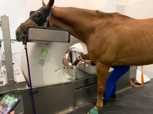
The answer came from computed tomography (CT).
When the Clinical Picture Doesn't Match the Radiographs
Navicular syndrome remains one of the most common causes of chronic forelimb lameness in horses, particularly in Warmbloods, Quarter Horses, and Thoroughbreds. Yet diagnosing it can be frustrating.
Many traditional diagnostic tools lack either sensitivity or specificity. Hoof testers may be negative even in confirmed cases, while palmar digital nerve blocks do not always completely resolve lameness1. Radiography remains the most commonly used imaging modality, but substantial bone density changes may be required before abnormalities become visible2.
This creates a familiar scenario in practice: a horse with convincing clinical signs but relatively unimpressive radiographs.
CT Reveals the Bigger Story
In this gelding, CT uncovered a far more extensive disease process than radiography suggested.
Beyond the enthesophyte seen on radiographs, CT identified1:
- Bone cysts within both navicular bones
- Marked sclerosis, particularly on the right side
- Enlarged vascular channels
- Additional enthesophytes at the distal and lateral aspects of the navicular bone
- Mineralization within the deep digital flexor tendon (DDFT)
- Focal abnormalities and irregular margins within the DDFT
These findings highlighted an important reality of navicular syndrome: the condition rarely involves the navicular bone alone.
Pain may originate from a combination of osseous and soft tissue lesions, making it difficult to fully characterize the disease using conventional imaging alone1,3.
Why Advanced Imaging Matters
For years, MRI has been considered the gold standard for evaluating horses with foot pain because of its superior soft tissue visualization.
However, CT is increasingly demonstrating significant clinical value.
Not only can CT identify characteristic navicular bone lesions such as sclerosis, cyst formation, enlarged vascular channels, and enthesophytes, but it can also reveal abnormalities within the DDFT and surrounding structures1.
An additional practical advantage is time. CT examinations can often be completed far more rapidly than MRI, reducing anesthesia duration while still providing detailed assessment of both bone and selected soft tissue structures1.
For horses with chronic, unexplained lameness, that efficiency can make advanced imaging more accessible in clinical practice.
Treatment Is Only as Good as the Diagnosis
Following CT evaluation, the horse received a multimodal treatment approach consisting of therapeutic shoeing, corrective trimming, and aseptic navicular bursa injection with triamcinolone and gentamicin.
The response was encouraging.
Within two months, appetite improved, body condition score increased from 3/9 to 5/9, and gait quality improved substantially1.
Yet perhaps the most important lesson was not the treatment itself.
It was the realization that radiography had revealed only a fraction of the pathology present within the foot.
The Take-Home Message
Navicular syndrome is rarely a simple disease. Its causes remain incompletely understood, with biomechanical factors, hoof conformation, and hereditary influences all believed to contribute to its development.
What is becoming increasingly clear is that relying solely on conventional diagnostics may underestimate the true extent of pathology.
When clinical signs persist despite treatment—or when radiographic findings fail to explain the severity of lameness—advanced imaging can uncover lesions that fundamentally change both prognosis and management.
Sometimes the challenge is not finding the navicular lesion.
It's discovering how many lesions are actually there.
References
- Lee S, Lee EB, Park KW, Jeong H, Kang TY, Seo JP. Computed tomographic findings of navicular syndrome in a horse. Journal of veterinary clinics. 2021;38(2):94-7. https://doi.org/10.17555/jvc.2021.04.38.2.94
- Harcourt M, Smith C, Bell R, Young A. Magnetic resonance and radiographic imaging of a case of bilateral bipartite navicular bones in a horse. Australian veterinary journal. 2018 Nov;96(11):464-9. https://espace.library.uq.edu.au/view/UQ:c67f677/UQc67f677_OA.pdf
- Frątczak M, Włodarek J, Frąckowiak H, Komosa M. Insight into the pathomorphology of the distal border of the equine navicular bone. Acta Veterinaria Brno. 2017 Jul 13;86(2):123-31. https://actavet.vfu.cz/media/pdf/actavet_2017086020123.pdf
Related Contents

Upcoming Event
ECG Interpretation Made Easy for Small Animal Practitioners
Electrocardiography (ECG) is an essential diagnostic tool in small animal practice, yet many clinici...
Upcoming Event
Diagnostic Imaging: Common Mistakes in Everyday Practice
Diagnostic imaging is an indispensable tool in veterinary practice, supporting the diagnosis of a wi...
 Subhasis Batabyal 2 (1).jpg)
Upcoming Event
Clinical Biochemistry and Veterinary Practice: Recent Trends in Veterinary Laboratory Diagnosis
Clinical biochemistry plays a pivotal role in modern veterinary diagnostics by providing objective i...

Upcoming Event
Emergency Drugs Every Veterinarian Should Know
Timely administration of the right emergency drug can make the difference between life and death in...

Upcoming Event
Obesity Management: Building Successful Weight Loss Programs in Dogs
Canine obesity is one of the most common nutritional disorders encountered in veterinary practice an...
.jpg)
Upcoming Event
Approach to Kidney and Liver Injury
Kidney and liver injuries require timely recognition and a structured diagnostic approach. Explore p...

Upcoming Event
CBC Interpretation in Small Animal Practice
A complete blood count (CBC) is one of the most valuable diagnostic tools in small animal practice....

Upcoming Event
Management of Fat and SNF in Milk of Dairy Animals
Milk fat and solids-not-fat (SNF) are key indicators of milk quality, nutritional value, and economi...