Article
FMD Diagnosis Challenges Every Veterinarian Should Recognize
Foot-and-mouth disease (FMD) remains one of the most important vesicular diseases affecting cloven-hoofed animals because of its rapid transmission, severe production losses, and major implications for animal movement and trade. One of the biggest challenges in field practice is that FMD can clinically resemble several other vesicular and erosive diseases, making early recognition extremely important for practicing veterinarians1.
For field veterinarians, the ability to identify suspicious lesions early and respond appropriately can significantly reduce the spread of infection within and between herds.
Recognizing the Classical Clinical Presentation
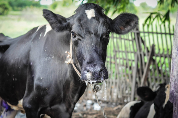
The classic presentation of FMD in cattle includes:
- Fever
- Hypersalivation
- Lameness
- Vesicles in the oral cavity
- Lesions on coronary bands
- Interdigital clefts
- Teats and muzzle lesions2
Vesicles initially appear as pale or blanched areas before progressing into fluid-filled lesions. These vesicles often rupture within 24 hours, leaving painful erosions and ulcers16. Animals may stop eating, show reluctance to move, or separate themselves from the herd because of pain associated with foot lesions.
In some cases, chronic hoof deformities may develop due to damage around the coronary band16. Secondary bacterial infections can further worsen recovery.
Young animals deserve special attention because sudden death due to viral-associated myocarditis may occur even before vesicular lesions become obvious1. This makes complete herd examination extremely important whenever unexplained mortality is reported.
Why Clinical Diagnosis Alone Is Risky
One of the most important practical points for veterinarians is that FMD cannot be confirmed based on clinical signs alone.
Several diseases can resemble FMD clinically, including:
- Vesicular stomatitis
- Bovine herpesvirus infections
- Bovine parapoxvirus infections
- Bovine viral diarrhea
- Malignant catarrhal fever
- Bluetongue-associated oral lesions3
Physical injuries such as chemical burns or thermal trauma may also mimic erosive lesions.
A useful field clue is lesion distribution and herd involvement. Vesicular diseases affecting multiple animals simultaneously, especially with lesions at multiple body sites, should always raise suspicion for FMD3. Rapid spread within a herd is another important warning sign.
Sample Collection: The Most Critical Step
For practicing veterinarians, proper sample selection directly affects diagnostic accuracy.
The best diagnostic samples include:
- Vesicular epithelium
- Vesicular fluid
- Oropharyngeal samples from clinically affected animals1
These samples contain high viral loads and are suitable for real-time RT-PCR and antigen ELISA testing1.
Blood samples are less useful during later stages because viremia is short-lived18. Serology becomes more useful approximately two weeks after infection when antibodies become detectable4.
When collecting samples:
- Select freshly ruptured vesicles whenever possible
- Avoid heavily contaminated lesions
- Maintain cold chain during transport
- Use appropriate biosecurity precautions
- Minimize farm-to-farm movement after suspect examinations
The Challenge of Subclinical and Carrier Animals
A major diagnostic complication is the presence of subclinical infection and persistent carrier animals.
Animals may shed virus before visible lesions develop1. In addition, approximately half of infected cattle may enter a persistent carrier state after recovery4,5,6. These animals may appear clinically normal but can harbor virus in the upper respiratory tract.
Routine oral swabs are often inadequate for identifying persistent infection24. Specialized collection using a probang cup is required1.
Vaccinated animals can also become subclinically infected and later develop persistent infection without obvious clinical signs1. This makes surveillance and monitoring more complicated in vaccinated populations.
Practical Field Recommendations for Veterinarians
Whenever vesicular disease is suspected:
- Isolate affected animals immediately
- Restrict unnecessary movement of animals, people, and equipment
- Conduct complete herd examinations
- Document lesion distribution carefully
- Use dedicated protective clothing and footwear
- Maintain strict disinfection practices
- Arrange rapid laboratory confirmation
Early suspicion and timely reporting remain the most important factors in outbreak control1.
Conclusion
Diagnosing FMD in field conditions requires more than simply identifying mouth or foot lesions. Practicing veterinarians must evaluate lesion distribution, transmission patterns, herd involvement, and disease progression together. Because several diseases can clinically resemble FMD, laboratory confirmation remains essential for definitive diagnosis.
Early recognition, proper sampling, strict biosecurity, and rapid response can significantly reduce disease spread and improve outbreak control outcomes. For veterinarians working in livestock practice, maintaining a high index of suspicion for vesicular diseases remains one of the most important responsibilities in protecting herd health.
References
- Arzt J, Sanderson MW, Stenfeldt C. Foot-and-mouth disease. Veterinary Clinics of North America: Food Animal Practice. 2024 Jul 1;40(2):191-203. https://www.sciencedirect.com/science/article/am/pii/S0749072024000033
- Stenfeldt C, Hartwig EJ, Smoliga GR, Palinski R, Silva EB, Bertram MR, Fish IH, Pauszek SJ, Arzt J. Contact challenge of cattle with foot-and-mouth disease virus validates the role of the nasopharyngeal epithelium as the site of primary and persistent infection. Msphere. 2018 Dec 26;3(6):10-128. https://journals.asm.org/doi/pdf/10.1128/msphere.00493-18
- Stenfeldt C, Eschbaumer M, Rekant SI, Pacheco JM, Smoliga GR, Hartwig EJ, Rodriguez LL, Arzt J. The foot-and-mouth disease carrier state divergence in cattle. Journal of virology. 2016 Jul 15;90(14):6344-64. https://journals.asm.org/doi/pdf/10.1128/jvi.00388-16
- Bertram MR, Yadav S, Stenfeldt C, Delgado A, Arzt J. Extinction dynamics of the foot-and-mouth disease virus carrier state under natural conditions. Frontiers in Veterinary Science. 2020 May 20;7:276. https://www.frontiersin.org/journals/veterinary-science/articles/10.3389/fvets.2020.00276/pdf
- Stenfeldt C, Arzt J. The carrier conundrum; a review of recent advances and persistent gaps regarding the carrier state of foot-and-mouth disease virus. Pathogens. 2020 Feb 28;9(3):167. https://www.mdpi.com/2076-0817/9/3/167
- Hayer SS, Ranjan R, Biswal JK, Subramaniam S, Mohapatra JK, Sharma GK, Rout M, Dash BB, Das B, Prusty BR, Sharma AK. Quantitative characteristics of the foot‐and‐mouth disease carrier state under natural conditions in India. Transboundary and emerging diseases. 2018 Feb;65(1):253-60. https://www.academia.edu/download/52151567/Hayer_et_al-2017-Carrier_Extinction_India.pdf
Related Contents
.jpg)
Upcoming Event
World Animal Day
World Animal Day highlights the importance of protecting animal health and welfare through responsib...
.jpg)
Video
Transjugular Removal of Canine Heartworms: A Clinical and Surgical Overview
Transjugular removal of canine heartworms is a specialized intervention used in selected dogs with s...

Article
Feline Pregnancy Endocrinology: Hormonal Changes and Placental Function
Pregnancy in cats involves a distinctive endocrine pattern in which ovarian and placental hormones w...

Article
Hormonal Control of Pregnancy Termination in Cats
Pregnancy maintenance in cats depends strongly on coordinated hormonal support, particularly progest...

Article
Feline Embryo Implantation and Early Placental Development
Pregnancy establishment in cats involves a sequence of precisely timed events between fertilization,...

Article
Ultrasonographic Monitoring of Pregnancy in Cats: A Gestational Timeline
Ultrasonography provides a practical way to follow the progression of feline pregnancy by identifyin...

Article
Feline Mismating: Pregnancy Diagnosis and Clinical Management
Mismating or unplanned mating is a common clinical problem in sexually mature queen cats because of...

Article
Medical Termination of Pregnancy in Queens: Drugs, Timing and Treatment Protocols
Medical termination of pregnancy (MTP) in queen cats should be approached according to the timing of...